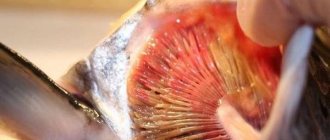
THE FISH HAS A COLOR CORNEA OF THE EYE
FSB
0
27 544
Share
Symptoms of the disease are clearly expressed - one or two eyes of the fish become cloudy, and sometimes the pupil increases in size.
The following circumstances may cause clouding of the cornea:
- Fungal or bacterial infection caused by dirty water in the aquarium or its overcrowding;
- Monotonous food and, as a consequence, the development of vitamin deficiency in fish. The disease is especially provoked by a lack of vitamin A in the body;
- Temporary clouding of the eyes, rarely but still occurring, is caused by chlorine contained in tap water if it has not been pre-settled before entering the aquarium;
- Eye fluke worms that attack the cornea from the inside, causing it to become cloudy;
- Eye injury;
- Cloudy eyes caused by old fish.
Clouding of the cornea of the eye can be caused by each of the above circumstances individually, or by their combined effect. From experience we can say that in many cases this disease is provoked by poor quality of water in the aquarium, as a result of which the fish’s immunity is weakened and any infection can cause an outbreak of diseases among all aquarium inhabitants.
Disease prevention
Simple aquarium prevention can significantly reduce the likelihood of corneal disease in fish. Conduct a daily inspection of the aquarium for any remaining fish food that was not eaten on time and remove any excess. Once a week, clean the soil using a siphon and replace about 1/3 of the aquarium water with fresh water. All these simple measures will help avoid the occurrence of this disease.
Treatment
First of all, you need to establish the cause of clouding of the cornea in fish. If the water in the aquarium has not been changed for a long time, then the reason in many cases lies precisely in this. Change the water daily in small portions and soon the disease will go away on its own and there will be no need to resort to treatment with various medications.
Due to the fact that constantly feeding fish with the same food can contribute to various diseases in fish, you should strive to make their menu as varied as possible. Feeding fish with high-quality fortified feed can give very good results. Currently, there is a large assortment of fortified dry food on the market, so there will be no problems purchasing them.
Tags: clouding of the cornea of the eyes, cloudy eye of a fish
Found an error or a dead link?
Select the problematic fragment with the mouse and press CTRL+ENTER. In the window that appears, describe the problem and send it to the resource Administration.
Causes of leukoma
An eyesore appears for a number of reasons:
- it may be congenital;
- Corneal leukoma can occur with age, when degenerative processes begin in the tissues and cells of the membrane;
- the development of leukoma may be influenced by viral or bacterial keratitis;
- The disease can be triggered by a corneal burn, eye injury, increased intraocular pressure, or ulcers on the cornea. Destruction of the epithelium is fraught with the formation of scar tissue;
- self-medication or incorrectly prescribed therapy for various eye diseases contributes to the appearance of cataracts;
- Leukoma can occur as a complication after surgery.
As a rule, the disease is accompanied by partial loss of vision, sometimes the formation of a cataract provokes complete blindness.
Types and severity of cataracts
White eyesore can be congenital or acquired. The second case is more common, since people are exposed to various lesions of the corneal layer, conjunctiva, and injure and burn their eyes. According to the severity, the anomaly is mild, moderate and severe. At first, barely noticeable spots appear that do not cause any discomfort to the eyes. With an average degree of damage, the spots thicken and become visually noticeable. The severe degree is characterized by the overgrowth of the spot with a vascular network.
Methods for diagnosing keratitis
It is possible to select an adequate treatment for keratitis only after determining the causes of the disease. The main diagnostic tool is the slit lamp: it is used to determine the depth, extent, and localization of inflammatory foci. If a bacterial, fungal or viral nature of the disease is suspected, bacteriological and cytological studies of smears from the surface of the cornea are prescribed. A fluorescein test may be prescribed to check the integrity of the epithelium, and an allergy test to identify allergens.
Symptoms and diagnosis
Leukoma often occurs without pronounced symptoms and obvious discomfort. As the disease progresses, symptoms become more pronounced:
- lacrimation increases;
- vision becomes blurred;
- the right and left eyes begin to see differently;
- eyes react poorly to bright light;
- Whitish spots form on the cornea.
As the corneal cataract develops, the spot turns yellow and degenerates into a fatty layer. If you suspect the appearance of this anomaly, be sure to consult an ophthalmologist. During the diagnosis, he will check visual acuity, assess the quality of refraction, analyze the condition of the cornea and fundus, and examine the anterior segment of the eye to determine the exact size of the cataract.
Treating eyesores
The method of treating a cataract directly depends on the cause of the disease, its form and stage of progression.
Medication method
This method is used in the primary stage of leukoma, when visual acuity is preserved. Most often, ophthalmologists prescribe drops and ointments that resolve scars, normalize metabolism and stimulate recovery processes.
Important: independent attempts to treat a cataract are prohibited!
Conservative-cosmetic method
The conservative-cosmetic method of treating leukoma is prescribed to those who have already formed a cataract, but does not have a particular effect on vision. In this case, colored lenses can be used to disguise the cosmetic defect. Just remember that they will not improve your vision.
Surgical method
The surgical method is considered the most reliable way to restore corneal function. To remove the damaged area of the cornea, the surgeon uses an electron microscope. He makes a small cut and removes scar tissue. Keratoplasty is often performed with a donor cornea transplant.
The operation can be end-to-end and layer-by-layer. In the first case, the clouded cornea is replaced with a new transparent one. In the second, only some of its layers are replaced, which is technically much more difficult to accomplish. The surgical method is used at the stage of already formed leukoma.
Remember that eyes require especially careful treatment. When they are normal, the cornea is completely transparent. If you notice even minor cloudiness, it is recommended to immediately contact a specialist. Any infection, eye damage or blurred vision is not advisable for self-medication.
Table of contents:
Causes of cyst manifestation
Where are eye cysts located?
Types of cysts Clinical symptoms Diagnosis of the disease Methods for removing an eye cyst Treatment of an eye cyst with a laser Procedure for laser removal Contraindications A benign tumor on the eyelid or mucous membrane containing fluid inside is called a cyst. Translated from Greek - “bubble”. It occurs in representatives of both sexes and all age categories. Quite often this formation is diagnosed in children. When it appears, you should definitely see a specialist to develop further treatment tactics. If left unattended, it can have a bad effect on the quality of vision.
If you do not contact the clinic to solve the problem, then:
- visual acuity decreases;
- eye mobility worsens;
- profuse lacrimation appears;
- frequent headaches occur;
- there is a possibility of developing into a malignant formation.
Laser removal procedure
- Local anesthesia in the affected area;
- an incision measuring 2 mm;
- a specialist inserts a laser source through the hole;
- the beam directionally evaporates the affected area and seals the blood vessels.
If the eyeball cyst is large or an abscess has appeared, then surgical removal under anesthesia is recommended. It takes place in several stages:
- limiting the surgical area with sterile materials;
- introduction of a staining solution to clearly define the boundaries of the lesion;
- fixation of the capsule;
- removal with a scalpel;
- rinsing the cavity with an antibacterial solution;
- connecting the incision using a continuous absorbable suture.
Rehabilitation is long and relapse is possible, unpleasant consequences such as hemorrhage, infection, suture dehiscence. In the postoperative period, antibiotics are prescribed, it is advised to reduce the load, and avoid contact with water. The applied bandage is not removed from the eye for five days. It serves as a barrier to dirt and bacteria. There are a number of restrictions on operations.
Methods for removing eye cysts
If you are diagnosed with an eye cyst, surgery will help radically solve the problem. Removing an eyeball cyst within healthy tissue is the most effective method of combating the formation.
- Cauterization with a laser beam is associated with the complete destruction of pathological cells. As a result, the likelihood of relapse is guaranteed to be low. The laser has a disinfecting and anti-inflammatory effect. Due to its undeniable advantages, if there are no contraindications, preference is given to this method. But if the formation is large, a significant number of laser pulses will be required, which will injure the surrounding tissue. Therefore, lasers with a shallow penetration depth are chosen, which makes it possible to remove only small formations, less than three millimeters in diameter.
- Surgical method.
Indications for surgical removal:
- medications do not have an effective effect;
- type of cyst;
- complicated neoplasms;
- pediatric dermoid cysts;
- a large or rapidly growing cyst.
Dermoid cysts do not respond to traditional treatment. This is a purely surgical diagnosis.
A cyst is a filled cavity measuring up to three millimeters or more. May consist of one or more chambers. When removing it, it is important not to disturb the integrity of the capsular bag, otherwise it will collapse, and this will complicate its excision. To clearly indicate its boundaries, markings were made on the outside. But the tear fluid blurs the designated boundaries. In addition, the tissues adjacent to the cyst swell under the influence of the anesthetic fluid, which also interferes with clear recognition of the boundaries. Therefore, to improve the visualization of boundaries, the following surgical technologies are used:
- Perform sequential aspiration of the cyst; based on the area of the collapsed section, it is determined whether it is single-chamber or multi-chamber;
- then the colored preparation is injected inside, giving the formation its previous shape;
- Local anesthesia is administered and the cyst is excised along clearly defined external boundaries.
With all the variety of existing types of neoplasms, only a specialist can choose the right treatment tactics if the patient is treated in a timely manner.