Symptoms
The symptoms of this disease are very diverse. Common symptoms for all types of fish: first, a sick fish loses its appetite, becomes lethargic, loses weight, its color turns pale, its fins are destroyed, its scales fall out; Sometimes there is darkening of the iris, bulging eyes, open ulcers, and black spots on the body. The listed signs can appear either singly or in combinations. Specific symptoms of the disease depend on the type of fish. In poeciliids, sick fish stay isolated from others and stop eating; exhaustion sets in, which is externally characterized by the fact that the back bends, the abdomen retracts, the eyes fall in, and bones protrude from under the skin and scales. In sick macropods, the skin is damaged and becomes red, the scales rise under the pressure of the liquid accumulated in the scale pockets and then fall out, sometimes an eyesore, bulging eyes occur, after which blindness occurs; the body becomes covered with black dots. In cockerels, the abdomen gradually increases, the skin begins to stretch and after 4-8 weeks becomes transparent; the fish become apathetic, but they move jerkily, often on their sides or belly up; stop eating. The zebrafish becomes bulging-eyed, and after some time the eyes fall out of their sockets, and the abdomen gradually increases in size.
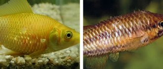
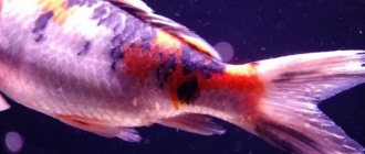
Photo: Fish tuberculosis, or Mycobacteriosis
Mycotic diseases of fish
Fish suffer from mycotic diseases due to the formation of fungi. They are multicellular or unicellular, non-chlorophyll-containing organisms belonging to lower plants.
Branchiomycosis
This is a fungus that affects the gill apparatus of fish. The causative agent of branchiomycosis is Branchiomyces demigrans and Branchiomyces sanguinis.
Causes. Fungi settle on the gill filaments. All fish species kept in improper conditions can suffer from the disease. The disease develops due to high water temperature and organic compounds of dead plants. The disease is developing rapidly.
Symptoms Sick fish do not have enough oxygen, pinpoint hemorrhages are visible on the gill leaves, and deformation of the gill covers occurs. The fish refuse food and swim at the surface of the water all the time, scooping up air with their mouths. Bright red and pale spots become visible on the gills.
Gill rot
Treatment. When the first signs appear, all fish are transferred to a quarantine tank and treated with malachite green oxalate, and the observation tank is cleaned and disinfected. Maintaining cleanliness and hygiene in the pool will help to avoid branchiomycosis.
Ichthyophonosis
A dangerous mycotic disease of pond and aquarium fish. Caused presumably by an imperfect fungus from the class Phycomycetes.
Causes. The causative agent is Ichtyophonus hoferi, which has a round or ovoid shape. A capsule is formed around the fungus, secreted by the affected organ. Hyphae are also observed in the form of blunt outgrowths that develop into an independent round body.
Symptoms The pathogen spreads hematogenously to various organs and tissues, where inflammation first develops, then encapsulation of the affected areas occurs. When functions are impaired, the fish stops responding to stimuli, its movement becomes erratic and sluggish. She stays off the coast. When the liver and kidneys are damaged, bulging eyes, ruffling of scales, and ascites are observed. Localization of the pathogen in the subcutaneous tissue, muscles and eyes leads to knobby swellings and ulcers, black spots on the skin.
Treatment. Not developed. But they must control the process of transporting fish. Be sure to feed fish with marine relatives only after heat treatment. Also, for preventive purposes, it is advisable to promptly disinfect ponds with quicklime or bleach.
Saprolegniosis ("cotton wool disease")
A mycotic disease of most fish species caused by opportunistic aquatic fungi from the class Oomycetes. More often this is a secondary disease; first, injured areas of the body or damaged eggs are affected, then the disease spreads to healthy areas and eggs.
Causes. The causative agents of the disease are representatives of the genera Achlya and Saprolegnia. The mycelium of these fungi is formed by hyphae having a limited number of transverse partitions.
Symptoms The most characteristic sign of the disease is cotton wool-like, fluffy white growths on the caudal and dorsal fins, head, olfactory pits, eyes and gills. Before the death of the fish, a loss of balance was noticed.
Treatment. In summer and autumn, for prevention, it is recommended to treat fish twice with basic violet K at the rate of 1 g per cubic meter. m of water for half an hour. 0.1% salt baths for 30 minutes are also suitable. To combat the disease, the water supplied to the hatchery is disinfected with ultraviolet rays.
Causes
Infectious disease. The causative agent is the bacterium Micobacterium piscum - a motionless thick rod 2-13 microns long. Affects the internal organs of fish. A large number of fish become sick at the same time. The most susceptible fish to the disease are: labyrinth, characin, carp, tropical cyprinids. Poeciliids, cichlids, and cetrarchs are less susceptible. It enters the aquarium from a reservoir where fish are found along with food, plants and soil, if it has not been boiled or calcined, as well as with fish, shellfish, plants, water and equipment from an infected aquarium. In addition, mosquitoes, ants, and cockroaches that crawl into the aquarium in search of water can be carriers of bacteria. Most often, the disease occurs in aquariums with poor conditions; one can even say that this disease is poorly maintained and the fish’s body is weakened. The pathogen is resistant to acid solutions and develops at a temperature of 18-25 degrees.
Proper sanitation for infected systems
Any porous materials, such as twigs or moss, should be thrown away. There is no way to effectively clean these materials. Fine granular substrate such as sand can be cleaned, but larger rocks and rocks have too many nooks and crannies for effective cleaning.
Because Mycobacterium spp have a special outer coating, choosing the right disinfectant is critical. Is a one percent Lysol solution most effective in eliminating systems previously infected by Mycobaterium spp.2? Never add Lysol to a fish system!
Treatment
At the very beginning of the disease, when the fish have not yet refused food, kanamycin 10 mg per 10 g of food is added to it. Effective treatment for subsequent stages of the disease has not been developed (The disease is incurable. For this reason, all therapy comes down to improving the conditions for keeping fish). Some mildly affected fish can recover on their own, provided that the conditions are optimal. Severely sick fish, other aquatic animals and plants are destroyed, the soil is boiled, the aquarium and equipment are disinfected with a 5% bleach solution or a 3% chloramine solution. To disinfect, pour 1 liter of solution into the aquarium and thoroughly wipe the internal and external walls and corners with a rag soaked in it several times a day. After this, the aquarium is rinsed several times, soil is filled in, and fresh, settled water is filled in, plants are planted, and fish are introduced a few days later.
Pathogens
Mycobacterium has unique properties; it is a large genus with more than 130 species that differ from each other and can affect not only animals, but also people. For example, obligate pathogens such as Mycobacterium tuberculosis (tuberculosis in humans) and Mycobacterium leprae, which becomes the causative agent of leprosy, are dangerous for humans. But saprophytes in the soil, Mycobacterium terrae, are also quite common and are absolutely safe.
Tuberculosis in fish is caused by the bacteria M. marinum, M. fortuitum, as well as M. Chelonae and some others. There are about twenty different species in total. At the same time, the bacterium affects more than 34 families and 150 species of aquatic organisms, but most often the disease develops in labyrinths, salmon, characins, cichlids and cyprinids, which are at risk.
Mycobacteria are rod-shaped, gram-positive microorganisms. Mycobacteria grow slowly. It causes a generalized infection in aquarium fish.
When mycobacteria penetrate the body of fish, granulomas form in the “victims” of microorganisms:
- in all internal organs;
- and in tissues (muscle, integumentary).
The mechanism of granuloma formation is not fully understood. It is believed that an important point is the absorption of mycobacteria by phagocytes. Moreover, in phagocytes the aggressor bacterium does not die, but multiplies. The body of a sick fish tries to localize the lesion. Connective tissue forms around the accumulation of phagocytes filled with mycobacteria. Replacement of organ tissue with connective tissue (fibrosis) leads to disruption of organ function and death of fish.
Mycobacteria are rod-shaped, gram-positive microorganisms. Mycobacteria grow slowly. It causes a generalized infection in aquarium fish.
Prevention
Mycobacteriosis is a typical disease of a weakened organism, which was facilitated by poor conditions for keeping fish, such as: poor sanitary and hygienic conditions, overcrowding, poor filtration, lack of oxygen. Therefore, the prevention of this kind of disease lies in creating impeccable, from a hygiene point of view, conditions for keeping fish in the aquarium. The level of microbes in the aquarium should be as low as possible. To do this, you can use ultraviolet lighting, which kills some germs. Important : These microbacteria can affect not only fish, but also humans. When working with an infected aquarium, you should be as careful as possible to avoid infection through the mouth (when sucking water through a hose with your mouth), through wounds or abrasions on the hands. After microbacteria enter the wound site after an incubation period (3 weeks), papular skin changes begin in these places. Spontaneous healing can occur after only 2 years. Therefore, you should immediately consult a dermatologist who will carry out treatment.
What is avian tuberculosis?
Avian tuberculosis is an infectious disease caused by bacteria, just like in humans. As in humans and cattle, this pathology affects exclusively parenchymal tissues, but not only the lungs, as in humans. The disease can be generalized in the liver, kidneys, intestines, glands, and genitourinary system. The bone marrow is also sometimes affected. As the disease develops, granulomas form in the tissues of the bird, in which bacteria accumulate.
This disease was first described in 1884, but it received the status of an independent disease with the name under which we know it now in 1890, after Koch conducted his research and discovered the “Koch bacillus” - the causative agent of tuberculosis.
The disease affects almost all varieties of poultry and poultry. Most often, chickens suffer from tuberculosis, less often - turkeys and ducks (moreover, wood ducks - more often), geese. The disease also affects flamingos, swans, pheasants, kites, ostriches, cranes, parrots and canaries. Among wild birds, pigeons, crows, and sparrows are susceptible to it. In total, about 80 species are affected, but pheasants suffer the disease the most severely. In chickens, young chicks are most susceptible to the disease.
Video
Mycobacteriosis tuberculosis
Mycobacteriosis or tuberculosis in aquarium fish.
How to disinfect an aquarium for tuberculosis in fish. Mycobacteriosis.
Identifying fish diseases! Their treatment. A guide for beginners. Lecture for aquarists.
Illustrations
photo 1. — Philomena, Red-eyed Moenkhausia (Moenkhausia sanctae-filomenae) sick with tuberculosis
photo 2 - Discus patient with tuberculosis
photo -3 spleen of a fish with tuberculosis, tubercles (granulomas, nodules containing a microorganism) are visible.
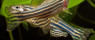
photo 4 - Pseudotropheus Sokolof, suffering from mycobacteriosis.
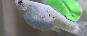
photo 5 - Lyalius patients with tuberculosis with a shrunken back, pale color and loss of scales.
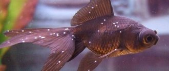
photo-6 - ruffled scales of a goldfish.
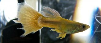
photo 7 - croaker guppy, curvature of the back due to tuberculosis
Diagnostics
Establishing an accurate diagnosis without microscopy is problematic. Because similar symptoms occur with aeromonosis (irritation of scales) and hexamitosis (appearance of dark spots on the body). Most likely, these diseases occur “paired” and both ailments need to be treated.
The disease can only be diagnosed after the death of the fish; for this, an autopsy is performed. During the study, tissue samples from the liver and other affected organs are taken, which makes it possible to identify the pathogen. The disease can only be determined indirectly by paying attention to the existing visual symptoms and disturbances in the behavior of aquatic organisms.