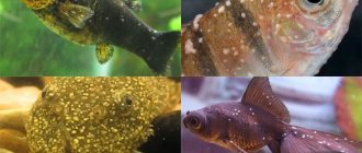
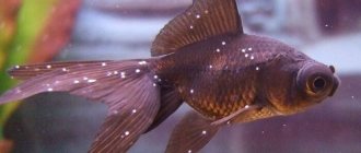
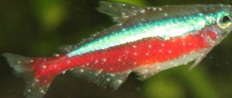
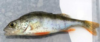
Ichthyophthiriasis/semolina is a contagious disease that affects the body of fish. The disease spreads very quickly throughout the entire aquarium. Therefore, if signs of the disease are detected, measures must be taken immediately.
The carrier of Ichthyophthyriosis is the ciliates Ichthyophthirius. These are the simplest biological organisms, reaching a size of 1 mm in adulthood. The parasites, once in the container, attach themselves to the body of the fish and begin to feed on the tissues of its body. As a result, white or gray grains are formed on the scales, reminiscent of semolina. Having sucked out all the vital juices of the victim, ichthyophthirius leaves the fish’s body and burrows into the aquarium soil. The infected fish dies, and Ichthyophthyriosis bacteria actively multiply and settle on the scales and gills of other inhabitants of the reservoir. If treatment is not taken promptly, all fish will die within one week.
There is also a tropical type of Ichthyophthyriasis. A distinctive feature of this variety is the development and reproduction of parasites directly on the body of the individual.
Causes of the disease
Semolina is an infectious disease caused by the ciliate bacterium. There are several ways it can get into the aquarium:
- Through live food from natural bodies of water (lakes, rivers, ponds, etc.);
- Through the ground;
- Through aquarium plants.
Fish can also become ill with ichthyophthyriasis when they are transplanted to another aquarium, when new fish are introduced, or when the conditions of keeping are changed. There have been cases when a store sold absolutely healthy fish, and the next day, after changing the aquarium, it developed semolina. This is due to the fact that bacteria already lived in the animal’s body, and the change in conditions had a beneficial effect on them and the ciliates began to multiply.
But when adding a new fish, it is important to pay attention not only to it, but also to the other inhabitants of the aquarium. After all, a newcomer can be a carrier of ciliated ciliates and, moreover, have some immunity to the parasite. Thus, the carrier fish does not get sick, but other fish that are not immune begin to become covered with white dots.
Prevention of Ichthyophthyriasis in a community aquarium
It is always easier to prevent the spread of a disease than to use difficult treatment methods later. Prevention will help save time and money by following simple rules when purchasing aquarium fish.
First, never purchase a pet from a pet store that appears sick or lethargic. Moreover, do not take a fish if it has new growths on its scales in the form of white or gray grains.
Secondly, do not buy aquarium fish whose neighbors seem to be infected. Even if the individual you like seems healthy, in the future there is a possibility that it will become infected with Ichthyophthyriasis.
Thirdly, after you have purchased the fish, quarantine it in a separate container for at least a week. Since sellers often catch fish for sale in dubious waters, there is a high risk of picking up a specimen infected with semolina. Quarantine will help determine whether the “newcomer” is infected.
If the worst does happen, aerate the container regularly during the early stages of infection. Good oxygen supply will help reduce suffering and prevent critical spread of infection.
Conclusion
Ichthyophthiriasis is a contagious disease that can cause a lot of headaches for aquarists. To reduce the risk of disease and reduce possible consequences, you need to clearly remember and follow the main rule: when buying a new fish, be sure to keep it in quarantine before placing it in a community aquarium. It must stay in a separate reservoir for at least one week.
If there is any suspicion of the spread of infection in the container, it is necessary to immediately take measures for prompt treatment. By following these recommendations, you can save the life and health of the inhabitants of the aquarium.
Watch the treatment video:
Share with friends on social media. networks
10% DISCOUNT FOR BUYERS FROM REGIONS OF RUSSIA AND CIS COUNTRIES FOR AQUARIUMS, FISH, PLANTS, ETC.
Symptoms
It is important to note that bacteria can parasitize a fish’s body for quite a long time and not show any signs of it. Therefore, you need to pay attention not only to the color of the scales, but also to the “hidden” symptoms.
- Initial stage of the disease. At this stage, the signal is the behavior of the fish. They begin to itch each other or the interior of the aquarium. This is how they try to get rid of irritation. Also at this stage (and subsequent others) there may be a deterioration in appetite.
- Then the fish begin to behave nervously and rush around the aquarium. Convulsions may occur. Infected animals lack oxygen, and therefore such fish stay on the surface of the aquarium, trying to capture air.
- The next stage of the disease is the appearance of characteristic white dots or tubercles on the body and fins of the fish. They can even be found in the mouth of an infected animal. Every day the number of these rashes increases, they begin to turn into ulcers and spread to healthy fish.
- The final stage of the disease is characterized by the loss of scales in animals. Loss of vision is also possible if the bacteria are localized in the visual organs. At this stage, it is almost impossible to save the fish.
Ichthyophthiriasis is extremely dangerous for fish, so when the first symptoms appear, treatment must be started immediately. Otherwise, as the disease progresses, it will be more and more difficult to cure the animal, and sometimes even impossible.
The causative agent of the disease, features of its development
its name “ ichthyophthyriosis ” to the pathogen. This is a ciliated ciliate - Ichthyophthirius multifiliis.
“Semolina” is one of the most common diseases of aquarium fish. This is explained by the rapid spread of the simplest organism in a closed aquatic environment. The single-celled ciliate easily penetrates the subcutaneous layer of fish, thanks to its miniature size and oval-streamlined shape.
Features of the structure of ciliates:
- The size of an adult organism does not exceed 1 mm.
- The entire surface is covered with numerous cilia, due to which it moves.
- In the middle of the body there is a large nucleus that looks like a horseshoe. It is called the nucleus - macronucleus. Near it is a small nucleus - micronucleus.
- Inside the transparent ifusoria, scattered vacuoles and lumps of food are collected.
- Scheme of the structure of the ciliated ciliate Ichthyophthirius multifiliis.
After being introduced into the skin of the fish, the ifusoria parasitizes there for about a week until it grows to the state of division. After which it sinks to the bottom and secretes a cyst containing a tomont . It is he who during the day is divided into numerous tomites , in the amount of 600 - 1000 pieces.
When they mature, they leave the cyst and become “vagrants” who infect a new victim. The duration of a full cycle is 7-10 days. That is why semolina spreads so quickly from sick fish to healthy individuals.
Treatment
It is easiest to cure ichthyophthyriasis in the initial stages, when the fish has not yet begun to become covered with white dots. In this case, the following tips will help:
- It is necessary to place the sick fish in a separate container to prevent infection of its neighbors in the aquarium.
- The aquarium in which the sick fish lived must be thoroughly cleaned. Completely replace the water, clean the soil, wash the interior of the aquarium (it is better to disinfect it by wiping it with a chlorhexidine solution) and install aeration.
- Add medications to water. This should be done not only in the aquarium where the sick fish is placed, but also in the general aquarium too. Because other fish may have a latent form of the disease.
The most effective recipes for medications for ichthyophthyriosis at home:
- 10 drops of iodine per 80-100 liters of water. Iodine does an excellent job of killing bacteria and disinfecting the environment. When adding iodine to the aquarium, the water temperature should be 26-28 degrees, no more.
- Malachite green dye is the most effective and safe method of treatment. When using it, the plants in the aquarium will not be harmed. The required concentration is 0.8 mg per liter of water. For an aquarium with a volume of 100 liters you will need 4 tablespoons of malachite green. It is not recommended to use this dye if there are fish without scales in the aquarium - it can cause irritation. If nothing else is at hand, then you need to reduce the concentration to 0.5 mg per liter of water. You need to add dye daily until recovery, while changing 25% of the water in the aquarium. To enhance the effect, you can add furatsilin at the rate of 1 tablet per 10 liters.
- 1 ml of 3% hydrogen peroxide per 10 liters of water. Add 2-3 times a day until the fish are completely healed.
There is also another way to treat ichthyophthyriosis. It consists in raising the temperature of the water in the infected aquarium to 32-33 degrees and adding table salt at the rate of 20 grams per liter. But this method of treatment is already outdated and may not be safe for fish. This is due to the fact that when this method was effective, there were only a few species of bacteria that could not withstand these conditions and died. Now there are much more modifications of this infection, and therefore the diversity of bacteria is higher. In some cases, especially when infected with a tropical type of ichthyophthyriosis, warm water and table salt can only improve the conditions for the reproduction of ciliated ciliates. Therefore, it is better not to use this method, risking the health and life of the animal.
What to do if spots appear on the fish
First of all, make sure that these spots are not a variant of the normal color of the fish. Sometimes such markings are present only in fish of one sex or appear during periods of maturation or sexual activity. However, there are many abnormal spots that can affect the condition of the fish. There are so many of them that we've grouped them by color to make them easier to reference. We started with light spots, among which there are those that are most common, namely white spots.
White or light spots
- White spots the size of a pinhead on the fish's head, body and fins are most likely the disease described in section 4.1.23. Cysts caused by Lymphocystis virus are less likely to occur (section 3.1.3). It is even less likely that they are caused by a protozoan parasite called Apisoma (section 4.1.1).
- Tiny whitish spots on the fins are usually tiny wounds, but sometimes they can look like the white spots described in the previous paragraph. Newly acquired fish that have these spots should be kept under observation.
- Whitish spots, which upon closer inspection turn out to be tiny tufts protruding from under the edges of the scales, may result from fungal infection of the fish (sections 3.3, 3.3.8).
- Small whitish tufts, especially on hard tissue (fins, gill covers), may represent Epistylis (section 4.1.5).
- Small whitish spots on poeciliaceae may indicate "guppy disease" (section 4.1.6).
- The whitish circles are likely wounds caused by leeches (section 4.2.6).
- A white spot that causes the lens of the eye to become opaque may be caused by eye flukes (section 4.2.5). See also clouding of the cornea (section 6.2) and section “What to do if?..”, No. 5.
- Irregular grey-white subcutaneous spots, which are commonly found in tetras (but also occur in some other fish) most likely indicate neon disease (section 4.1.13).
- Grayish or whitish spots of irregular shape are most likely caused by excessive mucus production (see section “What to do if...”, No. 19).
- Gray spots on the skin of the angelfish Pterophyllum spp. may be caused by Metrosporis parasites (section 4.1.8).
Note. During spawning, males of some genera of the cyprinid family (for example, barbs) develop white spots (tubercles) around the gills or head, and sometimes at the base of the pectoral fins. This is completely normal and is not a cause for concern.
Black or dark spots.
- Black spots on the body or fins are usually a sign of a disease called black spots (section 4.2.2).
- Black or dark spots around the mouth in East African cichlids is a disease called black chin (section 1.2.4).
- Dark or discolored, irregularly shaped spots on the body may represent superficial injuries (section 1.6.1), including burns.
Red spots
- Red spots on the skin may be wounds caused by large external parasites (ectoparasites), section 4.2, or subcutaneous hemorrhages resulting from systemic infection (sections 3.1, 3.2).
- Red spots or streaks on the fins (fin congestion) are often a sign of incipient fin rot (section 3.2.2).
Spots of other colors
- Tiny yellow-green spots, sometimes present on the body of the fish in such huge numbers that it seems as if it is completely covered with this color, are a sign of oodiniumosis (section 4.1.22).
- Tumors (section 6.7) may initially appear as spots. They come in all sizes and colors and are found anywhere on the body. (See section “What to do if?..”, No. 13).
Advice
When diagnosing one of the many different types of white or light spots, it is especially important to determine whether the fish is new or has been with you for a long time. It should be noted whether there are other signs of the disease present - for example, a fish with white spots itches, but a fish affected by lymphocystosis does not. The species to which the fish belongs is important (for example, cichlids and cyprinids do not get guppy disease or neon disease!), as well as the development and contagiousness of the disease (ichthyophthyriosis develops quickly, while others develop much more slowly). These differences are discussed in the relevant sections of Chapter 21.
What to do if your fish has tumors
Lumps, growths, or tumors on the surface of the body come in all kinds, shapes, sizes and colors. The range of possible causes is equally wide.
- Raised bumps (tissue damage) may result from certain bacterial infections (section 3.2) - for example, fish tuberculosis (3.2.3). These bumps may have a white or pale necrotic area (sometimes with an ulcerative pit) and an area of redness (hemorrhage).
- Digestive tract obstruction (including constipation (section 2.1)) sometimes causes a one-sided bulge, usually on the side or abdomen.
- An internal tumor (section 6.7) can also cause a similar bulge to appear. External swelling can appear literally anywhere on the fish's head or body. External tumors may be the same color as the surrounding skin, but are sometimes black (melanomas). They come in a variety of sizes and shapes and appear either singly or in clusters.
- Fish pox (section 3.1.2) initially causes irregularly shaped greyish or whitish spots, reminiscent of spots caused by excessive mucus production. These stains are soft at first, but over time they harden to a wax-like consistency. This disease is very rare in tropical fish.
- Encapsulated helminth larvae (sections 4.0, 4.2.2) located under the skin may appear as small growths on the body. There may be several such larvae or only one. They can have shades from light gray to dark gray if the fish itself is light.
- Whitish growths, usually forming clusters that resemble grapes or cauliflower in appearance and are especially noticeable on the fins, are manifestations of lymphocystis (section 3.1.1). (See section “What to do if...”, No. 12). Note: Some fish (especially males from the cichlid family) develop large fatty growths (nuchal humps) on their foreheads when they become adults. Sometimes these growths become permanent, and sometimes they shrink or disappear when the fish are not spawning. It is quite natural for mature females for one ovary to be more developed than the other and form an asymmetrical bulge on the body.
Advice
Tumors are more common in older fish. To distinguish a bump caused by an internal tumor from a bump caused by a gastrointestinal obstruction, determine whether the fish is excreting and eating (both of which are unlikely if the obstruction is severe). A lump formed by an internal tumor will develop more slowly.
What to do if the fish is too thin
Weight loss and exhaustion can occur for a variety of reasons.
- Direct pathogenic effect of the disease. Examples:
— pathogenic infection (usually systemic) caused by bacteria (section 3.2) or fungi (section 3.3). Happens rarely. Fish tuberculosis is a much more common cause of wasting (section 3.2.3).
— infection caused by protozoan endoparasites, such as neon disease (section 4.1.13) (occurs in tetras and some cyprinids);
disease associated with the formation of holes on the head (section 4.1.10) in cichlids; Heterosporis (section 4.1.8) in the angelfish Pterophyllum spp.
- A side effect of almost all diseases is that the fish does not eat because it is sick, and gradually becomes exhausted.
- Severe infestations of endoparasitic worms (sections 4.2.3, 4.2.4, 4.2.10, 4.2.12, 4.2.13) can also lead to wasting (as the parasites feed on their "host's" food). In this case, bloating may occur due to the huge mass of worms in the intestines. Some worms damage the intestinal lining and thereby affect nutrient intake.
- Poor nutrition or prolonged insufficient food intake, sections 2.0, 2.4 (see Chapter 7).
- Fish that stop eating while caring for their young (such as incubating eggs and fry in their mouths) can become very malnourished during this time.
- Spawning or childbirth can cause the female to suddenly lose weight. (See section “What to do if...”, No. 6).
Advice
“Too skinny fish” is a very relative concept. Most fish that have lived for some time in a home aquarium are rather more well-fed than nature intended. The fact that a certain fish is skinnier than others and has a flat belly profile does not necessarily mean that it is unhealthy in any way. This should not discourage the aquarist from purchasing such fish. True wasting (concave profile of the abdominal region) is a completely different matter. It does indicate poor health and inadequate food intake. If a fish that has been living in an aquarium for a long time suddenly begins to lose weight, this should be a cause for concern. The exceptions are when her diet or food intake has been deliberately reduced because she is overweight, or when she has recently laid eggs, or given birth, or is incubating eggs in her mouth.
What to do if your fish has growth retardation
Growth retardation - either permanent (the fish never reached the normal size for its species) or temporary (the fish grows slowly or stops growing for a while) - is a fairly common phenomenon. Some types of growth retardation (such as a genetic defect) cannot be cured, but others can be cured if the problem that caused the delay is addressed. The sooner this problem is resolved, the greater the chance of avoiding permanent stunting. Possible causes of stunted growth are listed below.
- A genetic defect (section 5.0), sometimes associated with inbreeding. It cannot be treated.
- Poor nutrition (sections 1.2.2, 2.4, 2.5) or insufficient feeding (section 2.4). See Chapter 7.
- Lack of appetite - see section “What to do if?..”, No. 6.
- Direct biochemical impact of unfavorable water parameters (sections 1.1, 1.2, 1.3).
- The direct result of disease caused by pathogenic organisms (section 3.0).
- A direct result of infestation by certain parasites, such as leeches (section 4.2.6) or intestinal worms (sections 4.2.10, 4.2.12, 4.2.13).
- Insufficient living space – the aquarium is overcrowded or too small for that particular fish.
- Hormones that suppress growth. Research has shown that there are a few species of fish (but further research may show that there are many more such species) in which the dominant (largest) individual in the brood produces hormones that suppress the growth of other fish. These hormones act only in close proximity and inhibit the growth of potential competitors of this individual - brothers and sisters. But fish that are in the same home aquarium, even the largest one, can be considered to be in close proximity!
Advice
Remember that in some species of fish, representatives of one sex may be larger than the other, and this is completely normal. There are fish whose growth rate is regulated by other factors. This dimorphism appears early and may become more pronounced over time as larger fish compete more successfully for food. Meanwhile, smaller fish often experience stress and loss of appetite and grow even more slowly because of this. As a result, large fish eat smaller ones. Therefore, young fish of some species must be regularly sorted by size and raised separately. When culling individuals from each brood, it is important to be aware of the possibility of early sexual dimorphism in size. Although culling obvious “dwarfs” is a useful thing, if all culling is carried out only by size, the matter may end up with only representatives of one sex remaining.
- Excessively frequent reproduction. This applies mainly to females, who devote a significant amount of energy to the formation of eggs. Females that incubate eggs in their mouths can suffer greatly because they do not eat at all during this time. Male fish of species that incubate eggs and fry in the mouth (either both parents or the father do this) may also suffer during this period. Other types of care for the offspring may also have a negative impact on the parent(s) guarding the eggs or fry. They cannot simultaneously fully care for their offspring and obtain food.
What to do if the fish has changed color
Changes in color in a fish are sometimes an indicator of changes in its health or the status it has in the aquarium (which can also affect its health). Fish that have noticeably darkened (or, conversely, lightened) may well be suffering from stress or illness. Abnormally bright colors can also indicate a problem.
Sudden or abnormal color changes should always be considered suspicious if they are accompanied by other general signs of illness.
The following color changes may be signs of specific diseases.
- If the fish is blind, it may acquire a persistent, solid dark color. This may be because the fish perceives the environment as complete darkness and therefore strives to conform to it (for the purpose of camouflage).
- Abnormally dark coloration is a very common sign of stress (section 1.5.2), but can also be seen in many other illnesses. It may reflect physiological changes or an attempt by the sick fish to become inconspicuous (a natural defense against predators and conflicts with other fish).
- An asymmetrical dark area on one side—usually the side of the head—may result from localized nerve damage inhibiting melanophore control. Possible causes are burn or mechanical injury (section 1.6.1), localized bacterial infection (section 3.2) (eg, abscess), or tumor (section 6.7). Permanent damage may result in permanent discoloration.
- Dark or discolored spots may result from burns or other superficial injuries (section 1.6. 1) - such as bruises.
- Black spots that expand over time (this happens over a period of days or weeks) are probably melanomas (section 6.7).
- In cichlids, dark areas around the mouth are a condition called black chin (section 1.2.5).
- In characins (less commonly in some cyprinids), fading of color is sometimes accompanied by the appearance of whitish or grayish spots under the skin - this is a sign of neon disease (section 4.1.13).
- Abnormally pale coloration may, among other things, indicate fish tuberculosis (section 3.2.3); shock (section 1.5.1); osmotic stress (sections 1.1.2, 1.6.2).
- Gray color (either the whole body or individual spots) may indicate excessive mucus production, which is a reaction to irritation caused by adverse environmental conditions (section 1.0) or parasites (section 4.0). It may also be a disease manifested as excessive production of skin mucus (section 4.1.18).
- A yellowish tint may be a sign of oodiniumosis (section 4.1.22).
- Reddened areas may be the result of damage caused by external parasites (section 4.1); injuries (section 1.6.1); irritation caused by acidosis or alkalosis (section 1.1.1), as well as ammonia (section 1.2.3); inflammation or hemorrhage due to systemic bacterial (section 3.2) or viral (section 3.1) infection; vitamin C deficiency (section 2.5).
- Large, pale pink areas on the abdomen are associated with dropsy (section 6.3) and some other systemic bacterial (section 3.2) or viral (section 3.1) infections.
- Discoloration of the fins (including the tail) along with signs such as lightened, grayish-white, frayed edges, reddened due to inflammation (there may be no redness), red streaks on the affected fin(s) may indicate fin rot (section 3.2 .2).
- Excessively bright or otherwise abnormal coloration may be a sign of damage to the central nervous system, resulting in loss of control of chromatophores. Possible causes are hypoxia (section 1.3.3), poisoning (section 1.2.1), acidosis or alkalosis (section 1.1.1), injury (section 1.6.1) or tumor (section 6.7).
Advice
To appreciate the significance of color changes, it is important to know what normal color changes a fish of a given type may exhibit. Many fish have relatively constant coloration, so any significant variation should be a cause for concern. However, in some fish, the color changes during their development and puberty. At the same time, there are fish that use color change as a means of communication and with its help demonstrate, among other things, their mood, social status, sexual status or courtship. Decoration and lighting of the aquarium can also play a role, as some fish become darker or, conversely, paler, trying to match their surroundings.
What to do if a substance resembling cotton wool appears on the fish
New growths that resemble cotton wool in appearance are usually a fungus, or cotton wool disease (section 3.3.3). Similar neoplasms occur in bacterial disease caused by the oral fungus Columnaris (section 3.2.4). The fungus usually affects the mouth area, but can also attack other parts of the fish's body, as well as the fins and gills.
What to do if the fish has holes
In addition to the mouth, gill slits and anus, fish have many other completely normal and natural openings. These are, in particular, the nostrils, which are located on the muzzle. Some fish have only one pair of nostrils, others have two pairs. In addition, fish have sensitive pores. These are tiny holes scattered throughout the head. There are one or more rows of the same holes - they go along the sides of the body and sometimes stretch all the way to the tail.
"Problem" holes.
- In cichlids, the sensitive pores on the head and lateral line (rarely) can become enlarged and infected due to a disease called perforation (section 4.1.10).
- If members of the cichlid family have enlarged or corroded pores, but there are no signs of pus in them, this may be a consequence of old age. There is no evidence that such pores cause any harm.
- Holes in the fins or body are usually injuries (section 1.6.1). The openings on the body may represent wounds left behind by external parasites such as Lernaea (section 4.2.1), leeches (section 4.2.6) or fish lice (section 4.2.7).
Advice
Aquarists who keep cichlids and have never seen head hole disease are aware of the threat this disease poses to their fish. They see nostrils and completely healthy sensitive pores and imagine that these are the first signs of this terrible disease. To avoid unnecessary stress for the aquarist and unnecessary treatment of healthy fish, we strongly recommend that any new cichlid keeper who is worried about developing this disease do the following. Let him ask a more experienced colleague to find and show him, as a standard, the normal holes that should be on the head of all cichlids.
DISEASES OF ZANIO PINK: DESCRIPTION, PREVENTION, TREATMENT, SYMPTOMS, PHOTOS
CICHLID DISEASES: DESCRIPTION, PHOTOS, TREATMENT, TYPES
HOW TO TREAT FISH WITH SALT IN THE AQUARIUM?
BARBUS DISEASES: SYMPTOMS, TREATMENT, PHOTOS
DISEASES OF COCKER FISH: EXTERNAL SIGNS AND TREATMENT
Special pharmaceutical preparations
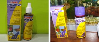
No matter how effective home remedies are to combat ichthyophthyriasis, drugs from the pharmacy are still more reliable and safer. In order to quickly and permanently cure fish, you must strictly follow the instructions of the drug. The following remedies are suitable for the treatment of ichthyophthyriasis:
- Antipar. Regulates the biochemical composition of water, killing harmful bacteria.
- SeraOmnisan. This drug is used only at the beginning of symptoms. At later stages it will be ineffective.
- AquariumPharmaceuticals.
- JBLPunktolULTRA.
All of these medications can be purchased at a pet store.
Immunity
Often fish get sick with semolina only once. Subsequently, they develop immunity to this disease. Therefore, if a carrier fish appears in the aquarium, then the recovered fish will either not react to the bacteria at all, or will suffer the disease in a very mild form.
Keeping fish healthy in an aquarium is not that difficult. The main thing is to be attentive to your pets so as not to miss the first signs of a serious illness and create comfortable conditions for their life. In this case, the fish will thank the owner with long life and harmony in the aquarium.
What is semolina
Semolina in aquarium fish, scientifically called “ichthyophthyriasis,” often affects even the most well-kept aquarium. The disease is infectious in nature and is caused by the ciliated ciliate parasite. The parasite affects: gills, fins, inner layers of skin. Externally, fish semolina looks like white tubercles or dots. Aquarists describe the disease this way: “the fish is covered with semolina.” These white dots are where the parasite accumulates. Ichthyophthiriasis proceeds aggressively, rapidly affecting every inhabitant. Treatment of semolina in aquarium inhabitants begins at the first signs of the disease, otherwise the fish die in less than a week. Semolina affects the immunity of fish, as a result of which pets are exposed to attack by other parasites and infections caused by bacteria and viruses. Often, when fish are affected by ichthythyriasis, other diseases are associated, so complex treatment is required. The most vulnerable small fish species and juveniles. They get sick and die first. But there is also a pleasant moment - this is resistance to the disease. Simply put, semolina in aquarium fish whose treatment was successful provides lasting immunity.
