Experienced owners know that aquarium fish, like other living creatures, can suddenly develop various diseases caused by improper care or the invasion of harmful organisms and infections into the pond. Most diseases can be successfully treated if the owner is able to correctly diagnose the aquarium fish during illness and take appropriate measures to save the pets. The sooner an aquarist comes to his senses, having noticed something wrong, the greater the chance of eliminating the disease of the aquarium fish, but for this you need to know the signs and symptoms of ailments, as well as their treatment at home.
Gill disease of unknown etiology (GILLD) or gill necrosis
Gill disease of carp is one of the most dangerous diseases that can cause mass mortality of farmed fish. The disease appears in early and mid-summer and subsides by autumn. Most often, two-year-old carp are affected.
The causative agent of the disease has not been identified. Gill disease of unknown etiology must be distinguished from branchiomycosis. The clinical signs of these diseases are largely similar. There are hypotheses about the viral and bacterial nature of the disease. It has been noted that the disease is often provoked by disturbances in the hydrochemical regime, primarily by an increased content of ammonia nitrogen and organic matter. Preventive measures aimed at creating favorable growing conditions are effective. It is recommended to use bleach or calcium hypochlorite, as well as quicklime. Quarantine restrictions are imposed on farms affected by gill disease. The disease can cause significant waste of live fish.
Saprolegniosis
Saprolegniosis is one of the most common fish diseases. It is believed that saprolegniosis is a secondary disease that occurs at the site of traumatic injuries on the fish’s body. In addition to trauma, saprolegniosis appears as a concomitant disease with other diseases, both infectious and invasive. The causative agent of the disease is lower fungi, mainly from the genus Saprolegnia, which are very widespread in nature. Saprolegniosis affects almost all freshwater fish that have been exposed to one or another effect or found themselves in unfavorable living conditions. Saprolegniosis often occurs in carp fish farms as a result of careless handling of fish, when kept in concrete cages, as a result of trauma during fishing, loading and unloading of live fish. The hyphae of the fungus penetrate the damaged tissues of the muscles, gills, and skin of fish, destroying the tissue. On the surface of the body, the fungus forms a coating similar to dirty cotton wool. Prevention is the main way to prevent saprolegniosis. All technological operations must prevent injury to fish. For preventive and therapeutic purposes, you can use drugs such as malachite green, brilliant green, and table salt. A type of saprolegniosis is Staff's disease. It manifests itself mainly in carp underyearlings during wintering. In this disease, fungi develop in the nasal cavities of fish. Fungal mycelium in the form of a cotton wool-like mass covers the head of fish, and fungal hyphae can grow into brain tissue. Staff's disease usually occurs in winter at very low water temperatures. Although there is evidence that this disease also occurs at temperatures of 5-6 degrees. Saprolegniosis of eggs is the scourge of hatcheries. Saprolegniosis affects the eggs of many fish species incubated in artificial conditions, mainly salmon and whitefish. The disease primarily affects dead eggs and quickly spreads to healthy eggs. Selection of dead eggs is a labor-intensive but effective method of preventing saprolegniosis. To combat this disease, malachite green, methylene blue, and brilliant green are widely used. Treatment of water with ultraviolet lamps and ozonation of water also prevent the development of saprolegniosis.
Types of aquarium fish diseases
There are as many types of diseases in aquarium fish as there are in other living creatures. The main causative agents of diseases are bacteria, viruses and fungi. The diseases themselves are divided into:
- Infectious,
- Invasive,
- Non-contact.
In the first and second cases, drug treatment is necessary, in the latter - improvement of the microclimate of the ecosystem. But it is important to remember that poor conditions in the aquarium will always affect the fish’s immunity and reduce it. And weak immunity is an entry point for infections and parasites.
A specialist must diagnose a particular disease. In large cities you can find an ichthyopathologist, but in the periphery you will have to cope on your own. The main thing is to notice problems in time and begin treatment. But the most effective way to successfully maintain an aquarium and fight diseases has been and remains prevention.
Branchiomycosis
An infectious disease of pond fish caused by a microscopic fungus. The causative agent Branchiomycosis destroys gill tissue. Fish with Branchiomycosis refuse to feed, accumulate near the shores, rise to the surface of the water, and assume a vertical position. The disease usually occurs in the summer, lasts 2-8 weeks and causes massive death of fish. Water bodies are quarantined, sick fish and corpses are caught and destroyed. In spring and autumn, ponds are disinfected with bleach or quicklime.
Mycotic diseases of fish
Fish suffer from mycotic diseases due to the formation of fungi. They are multicellular or unicellular, non-chlorophyll-containing organisms belonging to lower plants.
Branchiomycosis
This is a fungus that affects the gill apparatus of fish. The causative agent of branchiomycosis is Branchiomyces demigrans and Branchiomyces sanguinis.
Causes. Fungi settle on the gill filaments. All fish species kept in improper conditions can suffer from the disease. The disease develops due to high water temperature and organic compounds of dead plants. The disease is developing rapidly.
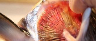
Symptoms. Sick fish do not have enough oxygen, pinpoint hemorrhages are visible on the gill leaves, and deformation of the gill covers occurs. The fish refuse food and swim at the surface of the water all the time, scooping up air with their mouths. Bright red and pale spots become visible on the gills.
Gill rot
Treatment. When the first signs appear, all fish are transferred to a quarantine tank and treated with malachite green oxalate, and the observation tank is cleaned and disinfected. Maintaining cleanliness and hygiene in the pool will help to avoid branchiomycosis.
Ichthyophonosis
A dangerous mycotic disease of pond and aquarium fish. Caused presumably by an imperfect fungus from the class Phycomycetes.
Causes. The causative agent is Ichtyophonus hoferi, which has a round or ovoid shape. A capsule is formed around the fungus, secreted by the affected organ. Hyphae are also observed in the form of blunt outgrowths that develop into an independent round body.
Symptoms. The pathogen spreads hematogenously to various organs and tissues, where inflammation first develops, then encapsulation of the affected areas occurs. When functions are impaired, the fish stops responding to stimuli, its movement becomes erratic and sluggish. She stays off the coast. When the liver and kidneys are damaged, bulging eyes, ruffling of scales, and ascites are observed. Localization of the pathogen in the subcutaneous tissue, muscles and eyes leads to knobby swellings and ulcers, black spots on the skin.
Treatment. Not developed. But they must control the process of transporting fish. Be sure to feed fish with marine relatives only after heat treatment. Also, for preventive purposes, it is advisable to promptly disinfect ponds with quicklime or bleach.
Saprolegniosis ("cotton wool disease")
A mycotic disease of most fish species caused by opportunistic aquatic fungi from the class Oomycetes. More often this is a secondary disease; first, injured areas of the body or damaged eggs are affected, then the disease spreads to healthy areas and eggs.
Causes. The causative agents of the disease are representatives of the genera Achlya and Saprolegnia. The mycelium of these fungi is formed by hyphae having a limited number of transverse partitions.
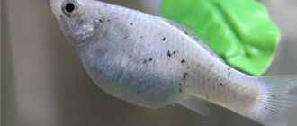
Symptoms. The most characteristic sign of the disease is cotton wool-like, fluffy white growths on the caudal and dorsal fins, head, olfactory pits, eyes and gills. Before the death of the fish, a loss of balance was noticed.
Treatment. In summer and autumn, for prevention, it is recommended to treat fish twice with basic violet K at the rate of 1 g per cubic meter. m of water for half an hour. 0.1% salt baths for 30 minutes are also suitable. To combat the disease, the water supplied to the hatchery is disinfected with ultraviolet rays.
Rubella of carp
Rubella (aeromonosis) of carp is one of the most common and dangerous diseases of carp and carp. In addition to these species, rubella is observed in crucian carp, tench, grass carp and some other species, but these cases are usually rare.
Rubella is a contagious disease, which, according to some researchers, is caused by a virus, while others believe it is caused by bacteria. There is objective evidence in favor of both hypotheses. Apparently, the term “rubella” covers several diseases with similar symptoms. Based on research conducted by the All-Union Institute of Experimental Veterinary Medicine, it was proposed to distinguish three independent diseases: aeromonosis caused by the bacteria Aeromonas punctata, pseudomonosis caused by bacteria of the genus Pseudomonas, and spring viral disease caused by the Rabdovirus cyprini virus (Vasilkov et al., 1978).
Rubella usually occurs in the spring or first half of summer. Sometimes the disease occurs in winter. Most often, two- and three-year-old carp are affected. The disease can cause massive waste. In the acute course of the disease, hemorrhages on the surface of the body, dropsy, bulging eyes, and ruffling of scales are noted. In the subacute course of the disease, the signs described above are supplemented by the formation of ulcers that have a whitish or reddish rim. The chronic form of rubella is most often observed in the second half of summer and is accompanied by the formation of ulcers on the skin and fins. The diagnosis can only be made by a specialist based on a set of studies.
To treat fish, drugs such as chloramphenicol, tetracycline, biomycin, methylene blue and a number of other drugs are used. In recent years, the use of probiotics has come into practice. Treatment is prescribed only by an ichthyopathologist, taking into account many factors relating to a particular body of water. Inappropriate use of antibiotics can cause negative results.
The only reliable method of rehabilitating a farm from rubella is summering, during which the ponds are drained, treated with disinfectants, and fish of all age groups are sold to the retail chain or disposed of. If sick fish live in a farm's water supply, it is almost impossible to improve its health.
Non-communicable diseases
Non-contagious diseases mean diseases that are not transmitted to other inhabitants of the aquarium. Most often, the development of ailments included in this category is caused by improper care or incorrect conditions. Treatment consists of eliminating the unfavorable factor or parasite that initiated the illness.
Alkalosis
Alkalosis, or alkaline disease of aquarium inhabitants, develops due to a too soft and acidic aquatic environment, the formation of which is facilitated by an abundant amount of vegetation and intense lighting. External signs of alkalosis are as follows:
- The color intensity decreases and the shine disappears.
- The fins spread out, the fish behave excitedly and unusually.
- Mucus is abundantly secreted from the gills.
If measures are not taken to carry out medical manipulations, the fish may go blind due to clouding of the eyes and die. In order not to torment yourself with the question of why the fish in the aquarium are dying, it is necessary to carry out work to save them in a timely manner. To treat alkalosis, you need to transplant sick pets into a container with a pH of 7-8.5, while simultaneously restoring the balance in the old aquarium.
Argulez
The development of argulosis is caused by the crustacean carpoede, or fish louse. These are quite large parasites with a rounded body shape, which are easy to notice upon visual inspection.
In addition, the following signs will indicate the appearance of argulosis:
- A large amount of bloody mucus from the wound due to the penetration of the parasite.
- The patient turns away from food, sways or rubs against hard surfaces.
Penetrating into the body, the carp eater destroys the skin and muscles, and releases a toxin into the body that poisons the fish. To save your pet, you need to remove the fish from the pond and use tweezers to pull out the parasite. After this, the wound is treated with a solution of potassium permanganate.
Acidemia
Acidemia, or ammonia poisoning, is observed among residents of resettled reservoirs, where there is a large concentration of waste in the aquatic environment. Symptoms of ammonia poisoning:
- The color of the scales darkens, the sick fish swims near the surface of the water.
- In rare cases, injuries to the gills are observed, and the fish tend to jump out of the container.
To treat victims, half the volume of water should be replaced, aeration should be increased, and the tank should be thoroughly cleaned of dirt.
Inflammation of the carp swim bladder (SVB)
The cause of the disease is not fully understood. The most substantiated point of view is the viral nature of the disease. The infection is transmitted mainly through direct contact between sick and healthy fish. Apparently, the pathogen can also be transmitted through water and soil.
Carp, carp and their hybrids are affected. Occasionally, cases of the disease have been reported in silver carp, pike, grass carp, crucian carp and pike. The main symptom of this disease is damage to the walls of the swim bladder. Inflammation often affects other internal organs. Fish of different age groups are susceptible to the disease. Fingerlings of carp suffering from STD usually die during wintering. In the acute form of the disease, massive waste is possible. Over time, fish develop immunity to this disease, and it gradually subsides. However, this is only possible if quarantine restrictions are observed. There are no specific drugs against this disease.
PARASITIC DISEASES
During ichthyopathological examination of fish living in natural reservoirs and in ponds of fish farms, many parasitic organisms belonging to different species are usually found. If the number of these organisms is small, the fish practically does not suffer from their presence. However, at high planting densities, characteristic of industrial growing conditions, the number of one or more species of parasites can increase sharply, leading to disease.
When growing carp, the most common invasive diseases are ichthyophthyriosis, trichodiniasis, costiosis, apiosomosis, phylometroidosis, bothriocephalosis, and caviosis. These diseases are usually treatable if diagnosed early.
Symptoms of diseases
Often, with certain diseases, certain symptoms arise, based on which it is possible to make a timely diagnosis and begin treatment of the disease in order to save the fish.
Bug-eyed
With this disease, the eyes become very swollen and often fall out completely. This symptom occurs during infectious infection, for example, with ichthyosporidosis, mycobacteriosis, etc. The method of treatment directly depends on this.
Causes. Bug eyes can occur due to infection of fish by viruses, bacteria or fungi. Along with this, provocateurs include physiological failures, trematodes, eye nematodes (worms), and lack of vitamins (beriberi).
Symptoms Cloudiness of the entire eye, the appearance of a whitish film, the eye moving away from the body. Advanced cases lead to the loss of one or both eyes.
Treatment. If bulging eyes are caused by a bacterial infection, it is initially treated using antibiotics and adding them to food. If a problem arises due to unsuitable living conditions, they begin by eliminating them: the water is regularly purified, the fish are fed in a balanced manner.
Bloating
Dropsy is accompanied by severe bloating and puffy scales. The fish becomes lethargic and breathes heavily.
Causes. Causes of abdominal enlargement include mycobacteriosis, aeromonosis, and nocardiosis. In addition to bacteria, a virus (spring viremia) can cause bloating. With gonadal cysts, an increase in the abdomen of females is also observed.
Symptoms A swollen abdomen, transparent skin due to a strong increase in the abdomen, curvature of the spine.
Treatment. The fish is immediately removed, observed and inspected. Fish are treated depending on the causes of abdominal enlargement, but most often the fish dies if the abdominal distension is caused by a bacterial infection.
If you learn to recognize the causes and symptoms of diseases, you will be able to prevent them or cope with ailments using effective methods. Following simple hygiene rules, frequent changes of water, and feeding fish with high-quality food will reduce the risk of infection in fish bred in natural or artificial conditions.
0
0
Copy link
Ichthyophthiriasis
This is one of the most dangerous ectoparasitic diseases that can cause massive waste of fish, especially juveniles. Almost all types of fish get sick. The disease is caused by ciliary infusuria, the name of which is translated from Latin as “Fish louse of many children.” The parasite develops and matures under the skin of the fish, and therefore it is resistant to many drugs that are effective for other diseases. Having reached maturity, the parasitic ciliate leaves the fish, sticks to underwater objects, and forms a cyst. After repeated division, several thousand daughter cells are formed in it. These cells then go out into the water and float freely for 2-3 days. If they manage to attach to the fish, they are embedded under the skin, where they develop.
Sick fish are weakened, stay in the upper layers of water, and react poorly to external stimuli. A small white rash similar to semolina is noticeable on the surface of the body and gills. The diagnosis is made only after a microscopic examination of scrapings from the surface of the skin and gills, since a “white rash” can appear with some myxosporidiosis, and can also be a manifestation of the “nuptial plumage” of male carp during the spawning period.
The fight is difficult because the parasite is located under the skin of the fish. Treatment is carried out only under the guidance of an ichthyopathologist. Prevention consists of preventing the entry of trash fish into reservoirs, pools, and cages where industrial cultivation is carried out. Transportation and transplantation of fish must be carried out using such preparations as malachite green, violet “K”, basic bright green, potassium permanganate.
Treatment: hexamitosis in fish (causes and symptoms)
The disease occurs when a flagellate parasite gets into the water, which penetrates the fish’s digestive organs and provokes the formation of cysts (Figure 3). The danger of this disease is that parasites can enter the aquatic environment along with feces.
The presence of the disease can be determined by its characteristic symptoms. At the initial stage, the individual’s skin color darkens, it prefers to stay away from the others, and at an advanced stage, white viscous discharge appears from the body.
Figure 3. Signs and treatment of hexamitosis
To treat hexamitosis, all infected individuals must be quarantined and antiparasitic drugs must be given. The remaining individuals also need to be given similar drugs, but not in therapeutic, but in prophylactic dosages. It often happens that the pathogen is immune to certain medications, so you need to try treatment with several medications one by one, or use them in combination.
Trienophorosis
The causative agent of the disease is a cestode from the genus Triaenophorus. Development occurs with the participation of two intermediate hosts: crustaceans and fish, mainly trout, perch, and a number of cyprinids. The main host is pike. If there are both trout and pike in the reservoir, the occurrence of this disease becomes very likely. The management of fish farms where fishing is organized and where trout are imported needs to implement a set of measures to prevent this disease. Treatment for the disease has not been developed. In order to prevent fish diseases in pond farms, it is necessary to protect the ponds from the penetration of pike into them from water supply sources.
With intensive infection, emaciation of fish, bloating of the abdomen, and pallor of the mucous membranes are observed. Deaths have been observed among fry and young of the year.
Philometroidosis of carp
Philometroidosis is caused by roundworms called nematodes. The main host of the parasite is carp, the intermediate host is cyclops. Female phylometras reach a length of 90 - 160 mm. They are localized in the carp's scale pockets in the area of the head, pectoral fins, and behind the gill covers. Males are much smaller than females and are usually found in the walls of the swim bladder.
When cleaning fish from scales, female phylometers look like pink-red rings with weak mobility. Although phylometra are not dangerous to humans, their detection in fish causes a negative reaction in the person preparing the fish. The buyer usually returns such fish to the sellers. The main cause of the disease is the transportation of fish from unfavorable fish farms to prosperous ones. Treatment of fish is possible using a number of drugs that have a high therapeutic effect.
Opisthorchiasis
Opisthorchiasis is a natural focal, severe helminthic disease of humans and carnivores, with primary damage to the liver, bile ducts, and pancreas. The causative agent is the Siberian or cat fluke (opisthorchis felineus). The parasite is small, 8-13 mm, oval in shape with two suckers. In the human body, the number of parasites can reach up to 40 thousand copies.
The lifespan of the parasite in the human body is up to 25 years. The cat fluke (opisthorchid) requires three hosts to develop.
The definitive host is humans, domestic animals (dogs, pigs, cats), wild animals (foxes, otters, arctic foxes) and other species of mammals whose diet includes fish. Eggs that are not dangerous to humans and animals are released from the intestines of the definitive hosts into the environment. Once in a pond, the eggs can remain viable for 5-6 months.
The intermediate host is mollusks. The eggs are swallowed by mollusks and, together with food, enter the gastrointestinal tract of the mollusk, where they turn into larvae. Mollusks hatch into thousands of larvae that float freely in the water, but are not dangerous to humans, so you cannot become infected with opisthorchiasis through water.
An additional host is carp fish. In fish, the larvae are localized in the muscles, eyes and other organs. When consuming fish infected with opisthorchid larvae, a person or animal becomes the definitive host.
Once in the body of the final host, the larvae, under the influence of gastric juice, are freed from the capsule shells, exit into the intestines and penetrate the liver, gallbladder, and pancreas, causing a severe disease. The accumulation of parasites in the bile ducts contributes to their blockage, disruption of the outflow of bile and deformation of the duct.
The disease can occur in acute or chronic form.
The acute form of the disease is caused by severe intoxication of the body and is characterized by increased body temperature, headache, malaise, swelling, rash, pain in muscles and joints. These symptoms occur 18-45 days after infection. In severe cases, the development of allergic myocarditis and eosinophilic small focal pneumonia is possible.
The chronic form of opisthorchiasis is characterized by symptoms of inflammation of the gallbladder (cholecystitis) with periodic exacerbations and remissions. Patients are concerned about heaviness and periodic pain in the right hypochondrium, epigastrium, loss of appetite, nausea, bloating, constipation or loose stools with the development of dysbacteriosis.
Bothriocephalosis
Bothriocephalosis is a helminthic disease of fish characterized by intestinal damage. Caused by the tapeworm Bothriocephalus acheilognathi from the family. Bothriocephalidae. Carp, carp, crucian carp, bream, bluegill, grass carp, silver carp, roach, ide, barbel, catfish and others are affected, but the most susceptible are the fry of carp, carp and grass carp, the infection rate of which reaches 80 - 100%. In this case, mass death of juveniles occurs.
Bothriocephalosis is widespread both in pond farms and in natural reservoirs. This is facilitated by uncontrolled transportation of fish, the presence of common sources of water supply, growing fish in head ponds, etc.
Fry and fingerlings are most intensively infected in July - August, when zooplankton develops abundantly in ponds and fish feed intensively.
Diplostomatosis
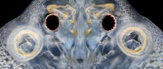
Diplostomatosis is an invasive fish disease caused by the larvae of digenetic flukes from the family Diplostomatidae, which parasitize the eyes of fish. Diplostomatosis is widespread among fish living in natural bodies of water. Juveniles and adult fish are sick.
Characteristic signs of the disease: clouding of the lens, formation of a cataract, blindness. Sometimes bulging eyes are observed. The membranes of the eyes become inflamed, impeding blood circulation, as a result the lens is destroyed, death and ulceration of the corneal tissue occurs, and the lens falls out. Saprolegnia fungi colonize the affected areas of the eye. Having lost vision, the fish does not take food well and loses weight; juveniles are stunted.
The causative agent of diplostomiasis develops with the help of two intermediate hosts: the first is a pond mollusk, the second is a fish. The main host is a fish-eating bird.
Columnaria: treatment
Columnaria is considered one of the most dangerous fish diseases, since its causative agent can be constantly present in a body of water, multiplying on organic debris (for example, on uneaten food).
Provoking factors for the development of columnarium are mechanical damage, lack of oxygen and elevated water temperature. At the initial stage of the lesion, the disease affects the skin and fins, gradually penetrating into the body. The disease can be detected by a simple external examination: characteristic light spots and stripes appear on the fins and skin, which without appropriate treatment can cause tissue necrosis (Figure 7).
Figure 7. Columnaria symptoms
Unfortunately, treatment of columnarosis is possible only in the early stages. To do this, antibiotics are used, which are simply dissolved in the pond water. However, in advanced stages, treatment will not be sufficiently effective, and the infected individual is simply destroyed.
Postdiplostomatosis
Postodiplostomosis is a common invasive fish disease, recorded both in natural reservoirs and in spawning and rearing and pond farms. It is characterized by damage to the skin, muscles, and curvature of the spine. It is manifested by the appearance of black spots of various sizes on the body of fish, from which the disease received its original name - black-spotted disease. These spots are formed as a result of the deposition of black pigment in the habitats of helminth larvae.
The disease is recorded mainly in water bodies in the southwestern regions of the country, where herons are more common. Infection of fish occurs mainly in the spring and summer, which is associated with the development of the pathogen. Various types of freshwater fish are susceptible to post-diplostomiasis - more than 60 species: carp, carp, bream, roach, grass carp, silver carp, rudd, sabrefish, roach, ram, silver bream, perch, white-eye, chub, podust, etc. Most of these fish species have commercial significance. The fry and fingerlings of these fish species are most sensitive to the disease. The body of the affected fry is deformed, the spine is bent, flexibility is lost, and growth slows down. Sick fish rise to the upper layers of the water, become weak and are easy to catch. Black spots on the skin form in various places: on the fins, gills, tail, back, abdomen, sides, cornea of the eyes, on the mucous membrane of the oral cavity, etc. The number of such spots is several tens or even hundreds.
Sources of invasion are infected fish, shellfish and herons that infest water bodies with helminth eggs.
Types of diseases
Diseases of aquarium fish are divided into several types depending on the cause that caused them.
Experts distinguish 4 main types:
- Bacterial;
- Fungal;
- Parasitic;
- Mechanical damage.
Treatment of diseases depends on the type and complexity of the course.
Bacterial
Bacterial diseases are caused by bacteria that are introduced into the aquarium by new pets, plants, or decorations.
- Aeromonosis. The disease is called rubella. Characterized by red stripes on the body and fins of fish. Associated with damage to the circulatory system by Aeromonas bacteria. In addition to red stripes, fish have bulging eyes, a swollen belly, and rapid breathing. The causes of the disease include poor water condition, high content of decomposing residues, nitrates, and lack of oxygen. There is no obvious cure for the disease. They use treatment of the aquarium, adding an antibiotic to the food in the initial stages.
- Columnaria. Bacterial infection is more typical for catfish and viviparous fish. It manifests itself as a fin disease in aquarium fish. They become shabby and torn. Ulcers appear on the body, and mucus appears on the gills, head and back. Behavior becomes tense and constrained. For treatment at the initial stage, a salt solution is used, in more severe cases - antibiotics.
- Dropsy. A bacterial disease associated with the accumulation of excess fluid in the body. Characterized by swelling of the body, protruding scales. Dropsy cannot be treated. The fish die over time. In the early stages, a folk remedy for diseases of aquarium fish is Epsom salt (1/8 teaspoon per 20 liters of water), as well as the drug Bicillin-5.
- Tuberculosis. A bacterial disease associated with the formation of nodules on internal organs, which form voids during decay. When the disease occurs, curvature of the spine occurs, ulcers form, and yellow nodules form in front of the eyes. Kanamycin and vitamin B-6 are used for treatment.
- Fin rot. The disease is associated with necrosis and complete disappearance of the fins. The disease is contagious, easily transmitted to other individuals, and without treatment leads to death. It is characterized by the appearance of milky-white areas on the edges of the fins and tail, a sloppy appearance, and exposure of the fin rays. For treatment, water changes are carried out and excess debris is removed. Specialized medications are used for antibacterial treatment. In advanced cases, antibiotics are used - levomycetin, chloromycetin.
- Bug-eyed. Eye diseases in aquarium fish are associated with protrusion of the eyeball above the surface of the head. The disease is not contagious and may disappear on its own. Telltale signs include a cloudy eyeball, a bulging, bloody or lacerated eye. For treatment, use Epsom salts, antibacterial drugs and antibiotics recommended for the treatment of fin rot.
Bacterial diseases also include cloudy eyes, swim bladder diseases, and mycobacteriosis.
Fungal
- Gill rot (branchiomycosis). The main pathogen is a fungus that settles on the gill filaments. The disease is contagious and is easily transmitted between individuals with weakened immune systems. Vivid signs are lack of appetite, changes in the size of the gill covers, apathy, and hemorrhages on the petals of the gills. Fungicides are used for treatment, and the aquarium is treated with antiseptics.
- Saprolegniosis. The disease is caused by the fungi Ahliya and Saprolegniacea. A characteristic feature is the appearance of white fluff on the body of the fish. As the disease progresses, the skin dies and ulcers form. Treatment is carried out with copper sulfate, formaldehyde, sodium salt or silver nitrate.
- Ichthyophonosis. Develops as a result of the fungus ichthyophonus. It is characterized by the appearance of deep ulcers in fish, the death of fins, and loss of coordination of movement in fish.
If aquarium fish diseases are diagnosed in a timely manner, then coping with the problem is much easier.
In the later stages, most diseases cannot be treated. Affected individuals should be removed. Rinse and disinfect the aquarium thoroughly.
Invasive
Invasive diseases in fish are caused by infection of individuals by helminths and arthropods.
- Lerneosis. The disease is associated with infection of fish by crustaceans from the group of copepods. Parasites enter the pet's body. The main location is the tail or dorsal fin. The defeat leads to difficulty breathing and the appearance of small white-green or red threads at the base of the fins. Treatment is carried out by bathing the affected individual in a bath with potassium permanganate and treating the aquarium with an antiseptic composition.
- Velvet disease. Capable of infecting all types of freshwater fish. The disease is caused by a single-celled dinoflagellate algae. Vivid symptoms are small nodules on the surface of the skin, anxious behavior, mucus secretion, the presence of a gray or yellow coating, and sticking of fins. Copper sulfate and UV rays are used for treatment.
- Glucose. One of the most dangerous diseases. Occurs as a result of infection by parasites of the genus Glugea. Signs include bulging eyes, moving on the side, and the appearance of blood-filled bumps in the whites that develop into ulcers. Glucose disease cannot be treated. After diagnosis, all fish and contents of the aquarium must be destroyed.
- Fish louse (carp eater, argulus). Associated with the defeat of individuals by tick-shaped crustaceans. The length of the parasite is up to 5 mm. A sign of the disease is the appearance of ulcers, inflamed areas, and red marks on the body. The parasite is noticeable upon careful examination of the individual. It occurs as a result of adding affected individuals to the aquarium or using water from an open reservoir. During treatment, parasites are removed using tweezers. The fish is kept for 30 minutes in a bath with potassium permanganate.
- Semolina. Associated with damage to the body by the parasites Ichthyophthirius multifiliis. They are attached to the fish’s body, fins, and gills. The disease manifests itself in the form of white spots, pinched fins, and loss of appetite. Pets become restless and constantly rub against objects. The cause of the disease is a decrease in water temperature and the introduction of parasites from the external environment. For treatment, it is necessary to increase the water temperature to 27 degrees and add quinine sulfate to the water.
In addition to the listed diseases, fish also have chylodonellosis, costiosis, trichodinosis, and parasite damage.
Related to violation of rules of care
Improper care of fish associated with violations of the temperature regime, feeding schedule, and cleaning of the aquarium can lead to the occurrence of non-infectious diseases.
- Temperature violation. Increased water temperature manifests itself in the form of overly energetic behavior of fish and oxygen deficiency. A low temperature leads to lethargy, colds, and death of individuals.
- Lack of oxygen. The disease is associated with a lack of oxygen, which is typical for overcrowded aquariums. It manifests itself in the form of restless behavior, gasping for air, lack of appetite, infertility, suffocation, and weakened immunity. To solve the problem, increase the volume of the aquarium and improve water aeration.
- Temperature violation. Increased water temperature manifests itself in the form of overly energetic behavior of fish and oxygen deficiency. A low temperature leads to lethargy, colds, and death of individuals.
- Chlorine poisoning. Occurs as a result of increased chlorine content in water. It is characterized by difficulty breathing, the formation of mucus on the gills, restless behavior, apathy, and a change in color to a lighter color. The problem is solved by immediately transplanting the pet into another container and replacing the water.
- Alkali disease. Characterized by increased or decreased pH levels. At elevated pH, fish exhibit spreading gills, a change in color to light, and the formation of mucus on the gills. In the case of a low pH, fearfulness, decreased energy, and movement sideways or belly up are pronounced.
- The appearance of neoplasms. Formations in fish appear as a result of ionizing radiation, oncogenic viruses, violation of the genetic code, and exposure to parasites. A characteristic sign is the appearance of visible neoplasms, a change in color to black, and swelling of the pet’s body.
- Obesity. Obesity in fish occurs when the fat content of food increases, poor diet, or overfeeding. Characterized by rounding of the sides, infertility, and apathy.
- Gas embolism. Gas embolism occurs as a result of oversaturation of water with oxygen. It is characterized by increased anxiety, immobility of the gill covers, the formation of bubbles on the surface of the fish, and sideways movement.
Treatment of aquarium fish diseases caused by poor care involves normalizing living conditions, replacing water and cleaning the aquarium.
Mechanical damage
Non-communicable diseases in fish arise as a result of mechanical damage.
The most common causes of injury include:
- Inappropriate neighborhood;
- Violation of fish care conditions;
- Sharp edges of decorations in the aquarium.
At the first sign of injury, the fish is removed from the tank and treated in a bath with potassium permanganate. All potentially dangerous items are removed or replaced with new ones.
Cavious
Cavious disease is a helminthic disease of carp, carp and their hybrids, black and white carp, characterized by intestinal damage. Caused by the cestode Khawia sinensis from the family. Caryophyllaidae.
The disease is recorded in all carp breeding zones. All age groups of fish are susceptible to the disease, but mainly fingerlings and two-year-old carp in the spring and summer. In nature, the pathogen persists in fish during the winter. Helminths are localized in the intestines and remain viable, and in the spring they begin to produce eggs. Cavia larvae remain in the body of intermediate hosts - oligochaetes, which also overwinter in water bodies. in the lower layer of soil. In the spring, when the ponds are filled with water, oligochaetes become active and rise to the top layer of soil. Fish, feeding at the bottom of reservoirs, eat infested oligochaetes and become infected with caviosis.
Symptoms Sick fish are inactive and their mobility is limited. they stay more in shallow water near the shores of the pond, their skin is dull. Nutritional status decreases and emaciation is noted. The gills and mucous membranes are anemic, there is swelling of the abdomen and redness of the anus.
Vasilenko V.V. Fishy odor disease / www.gastroscan.ru. 2021
| Authors: Vasilenko V.V. |
Fishy odor disease
V.V.
Vasilenko This disease interferes with participation in public life, complicates career growth, can deprive the main joy of life - live human communication, cause depression and even lead to suicide. Despair increases when doctors are unable to help, seeing before them a completely healthy person with ideal test results. A rare disease, and therefore little known to doctors, is called primary trimethylaminaciduria.
Time heals, but the doctor is faster. Proverb
The beginning of the history of this peculiar genetic disorder is lost in the past. In ancient legends one can find a description of trimethylaminaciduria in the hyperbolic spirit characteristic of myths. One of the legends says that the creator of the Indian epic "Mahabharata" - the divine sage Vyasa - was ugly, and such a pungent smell of rotting fish emanated from him that on his wedding night his first wife closed her eyes in horror, and therefore their son was born blind. Vyasa's other wife, sensing the smell, turned pale, and her son was always pale*. (* Ilyin G.F. Adaparva (First book) // Ancient Indian legend about the heroes of antiquity “Mahabhorata” - M.: USSR Academy of Sciences, 1958).
Slavic legends about mermaids living in water, precisely the female primary element of the four symbolic primary elements that make up the ancient Slavic world, are also inspired by this metabolic feature, because in women with a predisposition to trimethylaminaciduria, a peculiar odor appears or sharply intensifies on certain days of the lunar month (Fig. 1 ).
Rice.
1. Mermaid In Shakespeare’s play “The Tempest,” the jester Trinculo speaks of the wild and ugly slave Caliban, according to the author’s description: “What is this here? Man or fish? […] Fish; smells like fish; real aged fish stink. It looks like the cod is not the freshest. Outlandish fish! […] I decided wrongly, I don’t insist. This is not a fish, but an islander […]"
(translation by M.A. Kuzmin).
You can guess that the second name for trimethylaminaciduria is a disease with a fishy smell. No matter how surprising it may seem, the first scientific descriptions of this disease - excessive secretion of the odorous substance trimethylamine in people who are practically healthy in all respects - date back only to the second half of the 70s of the last century.
Trimethylamine is a very odorous substance. Some people feel its presence at a minimum concentration of 1:109! The figure is impressive, but in fact it is not so small: that is 10 trillion molecules of trimethylamine in a liter jar of air. Depending on the amount of the substance, different odors may be felt - from the characteristic smell of herring brine to the stench of decaying fish. (It is all the more surprising that in minute quantities trimethylamine emits a floral aroma!).
When receiving a referral for analysis, think about what you will do if the result turns out to be: a) positive, b) negative. If the answers coincide, there will be no need for analysis. Cochrane's aphorism
In the human body, trimethylamine is constantly formed, but normally it is inactivated, i.e. turns into an odorless substance. In primary trimethylamianaciduria, the enzyme that should carry out this transformation is missing, and trimethylamine is released in exhaled air and with all biological fluids (sweat, urine, genital secretions).
The severity of enzyme deficiency can vary, and therefore fishy odor disease expresses itself differently.
It usually appears in infancy and intensifies during puberty (sometimes later). In women, the symptom intensifies or occurs immediately before the onset of menstruation, as well as when taking birth control pills.
Stercus et urina medici sunt prandia prima. Exaliis paleas? Ex istis collige grana. - Feces, and urine, and sputum - doctors get a lot of money. This is the salt of the craft; other things are out of place to worry about. Character F. Rabelais
The genetics of the disease are as follows. Carriers of the defect (mutant allele) under normal conditions do not have a specific odor. If both parents have this defect, there is a high probability of having a child with fishy odor disease. Information about predisposition to trimethylaminaciduria is incomplete. However, overseas it is known to range from 0.5% in the UK to 11% in New Guinea.
Deficiency of an enzyme called flavin-containing monooxygenase 3 (FMO3) can be not only congenital - absolute or relative, but also acquired (malabsorption of nutrients and / or excessive proliferation of bacteria in the small intestine, chronic hepatitis, cirrhosis of the liver, renal failure).
Presumably the smell comes from those who are genetically predisposed to it.
Sometimes the enzyme is not enough due to increased formation of trimethylamine in the body.
Wisdom, like turtle soup, is not accessible to everyone. Kozma Prutkov
What to do for those whose body odor is very fishy? What measures can a person suffering from one of the forms of the disease with a fishy smell take on their own in order to smell almost like flowers?
- Avoid circumstances that cause increased sweating (physical activity, stress, strong emotions).
- Do not neglect personal hygiene products (soaps, body lotions) that are acidic (pH 5.5–6.5). Trimethylamine is an alkali; under moderate acidity conditions, only 0.02% of the original amount remains in a volatile, odorous form.
- Do not eat foods containing trimethylamine oxide, which is transformed by intestinal microbes into foul-smelling trimethylamine. These are sea animals: mollusks (oysters, mussels, squid, scallops) and crustaceans (crabs, shrimp, lobsters). Many types of mollusks and crustaceans are food for fish. Accordingly, saltwater fish must be treated with great caution. Moreover, infants with a mild form of trimethylaminaciduria begin to acquire a specific odor when their nursing mothers eat saltwater fish.
- Do not consume milk from wheat-fed cows: it contains significant amounts of the precursor trimethylamine.
- Avoid foods containing indole and its derivatives. Indole is an inhibitor of the FMO3 enzyme. It is found in plants of the cruciferous family, which include cabbage, radish, and rapeseed. Indole derivatives are included in dietary supplements that claim to ensure the health of the reproductive organs.
- In moderate forms of the disease, exclude products containing biochemical precursors of trimethylamine, primarily choline and lecithin (Table 1).
Table 1. Choline and lecithin content in selected foods (in mg per 100 g)
| Product | Kholin | Lecithin |
| Chicken egg yolk | 1713 | 9600 |
| Liver | 632 | 850 |
| Whole eggs | 566 | 3700 |
| Soy flour | 340 | 1480 |
| Dry peas | 263 | 900 |
| Beef | 95 | 1000 |
| Oat groats | 94 | — |
| Wheat | 92 | 375 |
| Rice | 88 | 110 |
| Cod | 87 | 1 |
| Low-fat cottage cheese | 74 | 2 |
| Buckwheat | 60 | 460 |
| Premium wheat flour | 52 | — |
| Processed cheese | 48 | — |
| Pasta | 45 | — |
| Corn flour | 37 | — |
| Wheat bread | 31 | 39 |
| Rye bread | 29 | 32 |
| Cabbage | 29 | 130 |
| Potato | 28 | — |
| Whole milk | 15 | 60 |
| Carrot | 11 | 104 |
| Chicken egg white | 0,5 | — |
| Yeast | — | 500 |
| Sunflower oil | — | 1000 |
| Cottonseed oil | — | 1750 |
| Rapeseed oil | — | — |
| Soybean oil | — | 2500 |
It is useful to know that significant amounts of lecithin contain dietary supplements advertised as fish oil or omega acids for the prevention of atherosclerosis, as well as all sorts of “health-improving” ingredients and products for athletes that have not passed state certification.
They usually contain a large amount of choline and lecithin. Pregnant women, however, cannot limit themselves to choline - it is needed for the formation of the fetal nervous system. For the biosynthesis of choline in the body, an essential component is required, i.e. The sulfur-containing amino acid methionine, obtained by the body only from the outside with food. There have been no studies of the effect of dietary methionine restriction in trimethylaminaciduria. Women should avoid using birth control pills, replacing them with other methods of contraception.
The hormones contained in these pills, which are produced in increased quantities during menstruation, increase the odor.
Bad body odor can come from a variety of causes, but discussing them is beyond the scope of this book.
Paradox: many of us tend to react violently to other people's scents, often without noticing our own. As Arkady Averchenko wrote, “the air smells of sulfur and spoiled relationships” (Klinkov’s words in the story “Pokhodtsev and Two Others”).
Activities other than those indicated above are rather within the competence of a knowledgeable physician. Nevertheless, we will list substances that can be formed in the body (usually in the mouth and intestines) and give a certain odor to the breath and “winds”.
This is hydrogen sulfide (the pungent smell of rotten eggs) and its salts:
- carbon disulfide (slight odor, but pungent);
- dimethyl sulfide (unpleasantly sweetish);
- methylpropyl sulfide, diallyl sulfide (garlic);
- allylpropyl disulfide (onion);
- mercaptans:
- methyl mercaptan (smell of rotten cabbage); - propyl mercaptan (unpleasantly caustic); - allyl mercaptan (garlic).
You can also name nitrogen-containing compounds:
- ammonia (unpleasant sweetish odor);
- dimethylamine (fish, ammonia);
- indole, ethylenediamine, cadaverine (nauseating cadaverous odor);
- putrescine (a sickening smell of rotten meat);
- skatole (stench, like a toilet).
These substances are clearly presented in Table. 2*.
Table 2. Substances with a bad odor*
| Chemical substance | Odor characteristics |
| Hydrogen sulfide (H2S) | Strong smell of stale eggs |
| Carbon disulfide (CS2) | Slightly caustic |
| Dimethyl sulfide (C2H6S) | Unpleasantly sweet |
| Methylpropyl sulfide (C4H10S) | Foul-smelling compound |
| Diallyl sulfide (C6H10S) | Garlic |
| Allylpropyl disulfide (C6H11S2)** | Onion |
| Methyl mercaptan (CH4S) | Acrid, stale cabbage smell |
| Propyl mercaptan (C3H8S) | Unpleasant, caustic |
| Allyl mercaptan (C3H6S) | The smell of garlic |
| Ammonium (H4N) | Unpleasantly sweet |
| Dimethylamine (C2H7N) | Fish, ammonia |
| Trimethylamine (C3H9N) | The smell of herring brine |
| Ethylenediamine (C2H8N2) | Ammoniacal |
| Cadaverine (C5H14N2) | Corpse smell (Latin cadaver - corpse; found in fly agaric, brewer's yeast, cheese) |
| Putrescine (C4H12N2) | The smell of rotten meat (Latin putrescere - to rot, decay) |
| Indole (C8H7N) | Foul-smelling compound |
| Skatol (C9H9N) | Strong fecal (Greek skor, genitive skatos - droppings, feces) |
* In minimal quantities, skatole has the smell of jasmine, so it was added to the composition of the Red Moscow perfume as an odor stabilizer. Everything should be in moderation! ** Interestingly, whole garlic heads and bulbs do not contain these compounds, but contain a lot of the amino acid cysteine. When garlic or onion is cut, this amino acid is converted by enzymes into odorous disulfides.
Rice.
2. Drawing by Marina Turovskaya, “Newspaper 24” In addition to the above, a deficiency of FMO3 leads to a disruption in the metabolism of the biogenic amine tyramine, a chronically elevated level of which in the blood is fraught with depression, migraine, and hypertension. Tyramine is found in noticeable quantities in long-aged hard cheeses and smoked sausages. Trimethylaminaciduria may occur in premature newborns who are fed formulas containing large amounts of choline. (You shouldn’t be scared: this is due to the immaturity of enzyme systems.).
Medical Latin synonyms for the term “pathological halitosis”: Foetor (Foetor) ex Ore and Foetor (Foetor) Oris (can also be translated as “stench from natural orifices”).
An experienced doctor differs from an inexperienced doctor in only one way: an experienced doctor prescribes one medicine for 10 diseases, and an inexperienced doctor prescribes 10 medicines for one disease. W. Osler
Often doctors are not sufficiently prepared to deal with this specific problem, and people suffering from bad breath or halitosis are forced to undergo a lot of haphazardly prescribed examinations.
In fact, for a diagnostic search, an experienced specialist only needs to carefully examine the patient and ask him a few questions:
- Is the smell constantly present or not?
- How long ago did the smell appear?
- Is its occurrence associated with certain foods?
- Is the odor coming from the nose, mouth, or both nose and mouth?
- What medications are you taking?
- Is the smell noticeable to others?
Two mercaptans - ethyl mercaptan and butyl selenomercaptan - were registered in the 1998 Guinness Book of Records as the most foul-smelling substances.
Each of them, when inhaled, resembles a mixture of rotting cabbage, garlic, onions, burnt toast and sewer gases (electronic library “Science and Technology”, 02/03/2002). The smell is so nauseating that there was even discussion about creating a so-called skunk bomb from their mixture to disperse street riots (National Geographic magazine: US Military Is Seeking Ultimate “Stink Bomb”).
V.V. Vasilenko. Sixty essays on digestion (recommendations from a gastroenterologist to patients) Back to section
Argulez
Argulosis affects most pond fish, primarily juveniles, whose waste from argulosis becomes widespread. The causative agent of the disease is the gill-tailed crustacean of the genus Argulus. The crustaceans have a round shape, their size is 4-8 mm. They parasitize on the surface of the body, piercing the skin of fish. Inflammation usually develops at the site of injury, which is subsequently complicated by infection. Argulus can be seen with the naked eye. It looks like a rounded, flat gelatinous body with a diameter of several millimeters, often mobile. Two dark dots are clearly visible - the compound eyes of the crustacean. The disease is often observed in farms that organize recreational fishing and use wild fish (perch, bream, pike, roach, etc.) for stocking. Prevention is the most reliable way to prevent this disease. Effective treatment is possible only with the use of chlorophos, karbofos, and organochlorine compounds, which have a negative effect on the reservoir.
Aeromonosis of fish: treatment
Aeromonosis is an infectious disease also known as rubella (Figure 8). The disease can occur in any breed, since the causative agents of the disease live in most fresh water bodies.
Figure 8. Signs of aeromonosis and its treatment
Aeromonosis can develop in acute and chronic forms. In the first case, the development of the pathology is so rapid that in a short time it can cause the death of the entire livestock. In the chronic course of the disease, fish become carriers of infection and can gradually cause the death of more and more individuals. Aeromonosis is considered a dangerous disease, therefore, in the farm where it was discovered, a quarantine must be declared, and the fish are given antimicrobial drugs.
Lerneosis
Lerneosis is an invasive fish disease caused by parasitic crustaceans of the genus Lernaca. It is observed when growing fish in ponds and aquariums. The causative agent of lerneosis is L. cyprinacea and l. elegans, parasitizes herbivorous fish with L. ctenopharyngodnis, in carnivores - l. esocina. The crustaceans settle on the skin, fins, nasal pits, eye sockets, mouth and gill cavities of fish.
With a population of 10-15 specimens/p.z. cause anxiety in fish, increased sliming of the integument, and a bluish or gray coating appears on the body of the fish. The diagnosis is made after microscopy of scrapings. Treatment is selected taking into account environmental conditions and the condition of the fish. There are several effective drugs, which include malachite green, brilliant green, violet “K”, potassium permanganate, table salt, formaldehyde solution, etc.
Trichodinosis, apiozomosis, costiosis, scyfidiasis, trichophriasis
The diseases are caused by parasitic ciliates that develop on the surface of the body and gills of almost all cultivated fish species. When there are 1-5 parasites in the microscope field of view (10x10), clinical signs of the disease usually do not appear. With a population of 10-15 specimens/p.z. cause anxiety in fish, increased sliming of the integument, and a bluish or gray coating appears on the body of the fish. The diagnosis is made after microscopy of scrapings. Treatment is selected taking into account environmental conditions and the condition of the fish. There are several effective drugs, which include malachite green, brilliant green, violet “K”, potassium permanganate, table salt, formaldehyde solution, etc.
Infectious and invasive diseases of aquarium fish
Infectious fish diseases cause serious damage to the entire aquarium. Often their symptoms are hidden or disguised as other diseases. The insidiousness of infectious diseases is that the aquatic environment contributes to the instant spread of the disease. You can lose all the inhabitants of an aquarium in just a week. The most common infections:
White-skinned
The bacterium Pseudomonas dermoalba enters the aquarium with new pets or with decorative items if they were used in an infected pond.
Symptoms of the disease:
- The tail peduncle and fins turn white,
- Fish that are red in color turn yellow
- The sick fish swims near the surface of the water, holding its dorsal fin above the water,
- The body of the fish takes a vertical position upside down.
- The pet is motionless (deadly stage)
The disease is dangerous because it affects not only the pet’s skin, but also penetrates deeper, affecting the nervous system. There is a treatment for this disease. But it is effective only at the first signs. If you miss the onset of the disease, the prognosis is extremely unfavorable. Without treatment, all pets die in less than a week.
In the early stages of the disease, absolutely all inhabitants of the home pond are moved to another container in which chloramphenicol is diluted in a proportion of 200 mg per 1 liter of water for 5 days. The soil and decorations are disinfected, and the plants will have to be destroyed.
The second option for first aid for victims of an aquarium epidemic is the use of ordinary table salt. It is diluted in the proportion of 1 tbsp. l. for 20 l.
The third option is the use of medications that can be purchased as prescribed by a specialist at a veterinary pharmacy. He will also give recommendations on the dosage of the drug.
Restarting the aquarium after fish illness and completion of treatment is mandatory in full.
Branchiomycosis
A fungal disease caused by fungi of the Branchiomyces sanguinis and Branchiomyces demigrans families. Among aquarists, this disease is called “gill rot.” The circulatory system is affected: the vessels become clogged with blood clots.
Fungus appears in the aquarium due to beginners and feeding with live food. The development of the disease is facilitated by dirty water and lack of maintenance of equipment.
The prognosis for the course of the disease is unfavorable. The death of pets without treatment will begin on 3-4 days. Branchymycosis is very contagious.
Symptoms:
- The appearance of red and dark stripes on the gills in the first days of the disease
- General lethargy
- Loss of appetite
- Photophobia,
- In the last stages of the disease, the gills acquire a “marbling” appearance: the presence of pink, gray and whitish streaks.
- Tissue death
Treatment should be started as soon as possible. To begin with, the pets are placed in a quarantine facility to take fungicidal baths. Copper sulfate, rivanol, nystatin, acrimet, methylene blue, sea salt, phenoxyethanol are used.
The aquarium itself is thoroughly disinfected: the soil is siphoned and washed, the water is replaced either completely or half, but with the obligatory addition of methylene blue. The water is intensively aerated.
As a preventative measure, a number of measures are carried out:
- Careful care of the aquarium,
- Quarantine for newcomers,
- Disinfection of live food,
Surviving pets take a long time to recover. The gills can take up to a year to heal. But the scars, unfortunately, will remain for life.
Hexamitosis (hole disease)
Disease of the gastrointestinal tract. Caused by the flagellate parasite (Hexamita salmonis). Most cichlids, astronotus and discus are susceptible to hexamitosis.
Symptoms of the disease:
- Digestive disorder
- Thread-like stool with mucous structure. May contain inclusions of undigested food,
- Lack of appetite,
- Bloating (not always observed),
- Weight loss, hunching, flattening of the abdomen,
- Ulceration of the body and head with the release of serous fluid,
- General deterioration in appearance.
Hexamitosis often becomes chronic, which significantly worsens the quality of life of fish. Chronic ulcers remain. Completely treated fish recover, and ulcers on the body heal without a trace.
For successful treatment it is necessary to improve living conditions. It is not necessary to plant fish, especially cichlids; the technique is designed to carry out the complex directly in a reservoir. First of all, a thermal procedure is carried out: the water temperature is gradually increased by 3 - 4 degrees. During this period, aeration is increased so that the fish do not lack oxygen. It is better to remove the plants, as such warm water can be destructive for them. This treatment method is strictly contraindicated for African cichlids.
After this manipulation, medications can be used. For drug intervention, erythromycin, metronidazole, and ciprofloxacin are used. Metronidazole is used as the main medicine. It is diluted in the proportion of 1 tablet (250 mg) per 35 liters of water. In severe cases, 250 mg per 15 l. At the same time, change the water. The treatment regimen is as follows: the medicine is administered daily for 3 days, replacing 25% of water. Then every other day, and the volume of replacement is halved. In the second case, with extensive damage to the aquarium, the volume of replacement increases by 50%. The duration of treatment is 2 weeks.
Gyrodactylosis
Many fish diseases are invasive. Gyrodactylosis is one of them. Caused by the fluke parasite Gyrodactylus. Affects skin, gills, fins.
The main sign of the presence of a parasite is the appearance of a gray coating, reminiscent of mucus, on the gills and fins. In advanced cases, the destruction of the fins begins. Ulcers appear on the fish's body. The behavior of the fish changes: they try to swim as close to the surface of the water as possible, pressing their fins to their body. It seems that the fish is itching, as it constantly rubs its body against the decorations, as if trying to scratch itself. Gradually, the pets lose their appetite.
Treatment is carried out in a common aquarium using solutions of bicillin and azipyrine. In quarantine, you can use a solution of table salt and formaldehyde. It would be a good idea to add copper sulfate or methylene blue. The parasite is afraid of rising water, but with this method one should take into account the peculiarities of keeping aquarium fish
Glucose
Infestation by the sporozoan (Microsporidia), which causes glucose, occurs when water is ingested. In aquarium fish, the sporozoan infects the gills, eyes, and entrails.
The first symptoms of the disease appear acutely and immediately attract attention. The fish swims on its side, the body is covered with large tumors that rupture. The tumors are whitish and bloody in color. The fish develops bulging eyes.
There is no treatment for glutenosis in aquarium fish. Because of this disease, the fish are destroyed, including the plants, and the aquarium is completely restarted, thoroughly treating the soil, decorations, equipment, and the container itself.
Fin rot
The disease occurs after the fish are hypothermic. Most often those who suffer are those whom nature has awarded with veiled tails and fins. The fish are attacked by Pseudomonas bacillus. One of the diseases of aquarium fish is difficult to diagnose.
Signs that novices do not pay attention to, but are well known to experienced aquarists: cloudy fin colors that take on a whitish-blue tint. Sometimes the cloudiness affects the eyes. At the next stage, the fins begin to rot, sometimes to the point of complete self-amputation. If this happens, the pet dies.
Antipar bactericides, bicillin, and malachite green come to the rescue.
Dactylogyrosis
The disease is caused by the parasite Dactylogyrus. The disease affects the gills. Dactylogyrosis masquerades as branchiomycosis.
A sick fish tries to swim at the water's edge, constantly itches against decorations and equipment, breathes quickly, and loses its appetite. The fish's gills become pale or marbled, they stick together, the edges become torn, and a large amount of mucus appears.
For dactylogyrosis, two treatment options are allowed: in a general aquarium or in a fish tank. First of all, increase the water temperature if the type of aquarium fish allows this. They can be divided into different containers. Then some people increase the temperature of the water and add medications, while others are given only drugs. Bicillin, table salt and formaldehyde will help defeat the disease.
Dermatomycosis or saprolegnia
If the aquarium is poorly maintained, the mold fungus Saprolegniales may begin to actively develop in it. Fish's skin and gills are affected. Internal organs are rarely affected, mainly in the last stage of the disease. Dermatomycosis is a concomitant disease. Most often it occurs against the background of something else. In this case, solving the main problem leads to a cure for saprolegnia.
An attentive owner notices that white threads appear on the fish’s body. This is dermatomycosis.
Ichthyosporidiosis
A terrible disease that destroys the entire livestock. Belongs to fungi. The causative agent is Ichthyosporidium hoferi. All internal organs are affected. Bone and cartilage tissue are not affected, but this does not save the situation.
The symptoms are obvious and attract attention. The fish swim in jerks, constantly rub against various surfaces, often lie on their sides, and do not take food. The eyes become bulging, cataracts appear on them, the mouth is open, the body becomes covered with ulcers. The tumor process begins in fish. Organs are gradually rendered.
Ichthyosporidiosis is one of those diseases that has no cure. The entire aquarium is destroyed.
Alkalosis or alkaline disease
This disease does not have an infectious or parasitic etymology. Occurs against the background of errors in the maintenance of the aquarium. The cause of the disease is water, lighting and plants. What happens is that too much light and too much vegetation lead to acidification and increased softness. But, despite the fact that the disease is not infectious, it is just as dangerous and can lead to the death of pets.
Alkaline disease of aquarium fish is clearly manifested externally. The scales become dull, mucus appears on the gills, and the fish spread their fins. In the final stages, the fish lose their vision. Alkalosis forces them to quickly leave the aquarium, mainly at night. Therefore, if the owner finds a pet on the floor, he should immediately pay attention to the pH level.
All treatment for alkalosis involves calculating the pH level, which is carried out gradually outside the presence of the inhabitants. They should be placed in another container.
Asphyxia or suffocation
The fish are simply suffocating. This happens to careless owners who do not consider it necessary to purchase an aerator. The second reason is the irrepressible greed of aquarium owners, who stuff unfortunate fish like sprat into a tin can. As a result of overpopulation, there is a catastrophic lack of oxygen in the aquarium. Massive fish deaths begin. Treatment methods are obvious: resettlement of the inhabitants, increased aeration, total cleaning of the reservoir.
Acidemia or ammonia poisoning
The second disease that threatens fish as a result of improper actions by the owner is acidemia. Dirty water and soil, rotting food and feces, dead plants, rare water changes, all this leads to high concentrations of ammonia.
You want to escape from such a house, which is what the inhabitants do, jumping out of the water en masse. At the same time, the color of the fish darkens, they breathe frequently and heavily, and swim at the surface of the water. Of course, such behavior may indicate other diseases, but usually the owner understands that his mistakes in maintaining the home pond are the real reason.
Acidosis or acid disease
Sometimes the spawning of fish or the appearance of new ones leads to a decrease in the acid-base balance. Pisces become lethargic and sleepy. They swim lying on their side or completely upside down. Dead individuals are curled up in a ring. If the pH drops sharply, pets rush chaotically around the aquarium, jump out of the water, and mucus is released in large quantities. Adding baking soda, which will increase the acidity level, saves the situation.
Chlorine poisoning
A common mistake of lazy or illiterate owners. Chlorine poisoning occurs when fish are placed in tap water. The signs are the same as for suffocation. In principle, the process is the same - the fish suffocates. To normalize the condition of the pets, it is enough to transfer them to good water, which has been left to settle for two days. If there is no such water, then you can get rid of chlorine by heating the water to the boiling point.
In order to minimize diseases of aquarium fish, it is necessary to control many water parameters, chemical compounds, and the number of pets. If aquarium fish are kept correctly, they will not be afraid of diseases.
NUTRITIONAL DISEASES
| In industrial aquaculture with the transition to highly intensive forms of fish farming (in cages, pools, etc.), nutritional diseases cause damage. They are divided into 2 groups. The first includes diseases associated with the use of unbalanced feed in terms of fat, protein, carbohydrate, mineral and vitamin composition. The second group includes diseases that occur in fish as a result of consuming low-quality feed, contaminated with microorganisms (bacteria or fungi), their metabolic products, or containing oxidized fats. Nutritional diseases occur in fish of different species and ages. They reduce the growth rate of fish and can cause their death. The peculiarities of their manifestation require not only general, but also special approaches to their prevention. |
Prevention of helminthic diseases
- cook the fish for 15-25 minutes;
- cooking cutlets, meatballs, etc. - 15-25 minutes;
- baking pies with fish for at least 45-60 minutes;
- cold smoking of fish must be carried out either after pre-salting for 2-3 days, or after freezing;
- fry large pieces of fish spread out and always in fat for at least 20 minutes, fry small fish whole for 15-20 minutes;
- salt the caviar (at a temperature of 5-6°C), the ratio of the amount of salt is 6% to the weight of the caviar for 12 hours, for example: 60 g of salt per 1 kg of caviar, or salting the caviar in a 5% solution (50 g of salt per 1 kg of caviar ) at least 2 days with periodic mixing of caviar;
- salt: small fish for 14 days, large fish (over 25 cm) for 40 days with the addition of 2 kg of salt per 10 kg of fish;
- drying of fish for at least 3 weeks with preliminary salting for 2-3 days; d freezing: at 40°C-7 hours, at 35°C-14 hours, -28°C-32 hours.
The danger of contracting opisthorchiasis and diphyllobothriasis does not disappear all year round.