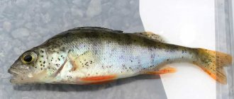
Argulus is one of 4 genera belonging to the class Branchiura (carpoeds or fish lice), phylum Arthropoda (Arthropods), subphylum Crustacea (Crustaceans). Carp eaters are characterized by a dorsoventrally flattened body and the presence of carapace lobes, which may or may not cover the biramous pectoral limbs. Individuals have a pair of compound eyes, four pairs of swimming limbs and an unsegmented abdomen. Argulus is a dangerous pathogen for fish living in the wild or in aquaculture. Once attached to a prey, the parasites appear as milky white or gray spots on the dark body of the fish; their eyes are always visible.
Pathogen
Argulus have horseshoe-shaped head shields covering the carapace lobes. The shape of the carapace varies depending on the species. In A. coregoni, the carapace lobes cover only the first three pairs of limbs, while in A. japonicus they cover all four pairs of limbs. At the junction of the carapace lobes and the head shields, a small notch is observed.
On the ventral side of the carapace there are two pairs of respiratory areas, which are species specific and serve for species identification. They have clear outlines and, unlike the rest of the carapace, are covered with a thin cuticle. There are five pairs of limbs on the cephalothorax. The first and second pairs are represented by antennules and antennae, respectively. The number and shape of spines and hooks covering these limbs are species-specific. The third pair is the maxillae or suckers, consisting of sclerite rods. The number of sticks varies depending on the type of carp eater. The fourth pair is represented by the mandibles, which include the proboscis or oral tubule, a structure located posterior to the preoral spine between the suckers. Behind the suckers is the next pair of limbs, that is, the maxilla. They are also characterized by the species-specific shape of the basal plates, the number of spines and scales. On the chest of the parasite you can see four pairs of two-branched swimming legs. In males, the legs additionally serve as a copulatory organ, which differs morphologically in different species. The posterior segment of the thallus is the abdomen [5].
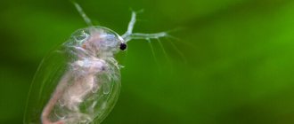
The illustration shows the head section of a carp eater (Magnification 60x. Wim van Egmond. Micropolitan Museum Rotterdam, the Netherlands).
A. coregoni is twice the size of A. foliaceus and has pointed rather than rounded ventral lobes. This species is very similar to A. foliaceus and can only be identified under a microscope.
A. foliaceus usually inhabits bodies of water rich in organic matter and is tolerant of salinities above 8-12 and temperatures above 25°C. Typically, A. coregoni infects fish in rivers, streams and cold, oligotrophic lakes with strong currents.
The parasite pierces the host's skin with a thin stylet and feeds on blood and skin using its proboscis and mandibles [3].
In addition to the harm that the carp eater itself causes, by feeding on fish tissues, it opens the gates for bacterial infections. In particular, it has been proven that F. columnare bacteria accompany infection with Argulus coregoni and increase the mortality of the host. In dead fish, areas of necrosis are observed on the skin around the tail and dorsal fin, which are typical signs of columnar blight.
The occurrence of a secondary infection is associated, on the one hand, with a violation of the integrity of the skin and mucus, and, on the other, with stress and a decrease in the immune status of the body [1].
Severe carp infestation on the skin of koi carp (news.ifas.ufl.edu/) increases by click Severe carp infestation on the skin of koi carp (news.ifas.ufl.edu/) increases by click
Subclass Mystacocarida
These are microscopic (0.5-1 mm long) maxillopods that live between grains of sand on sea beaches and in the sublittoral zone. Only 12 species of mystacocarids are known, but under favorable conditions, one cubic meter of sand can contain up to 15 million individuals. They are distributed on the coasts of Australia, Chile, South Africa, North America and the Mediterranean Sea.
The head is divided into 2 parts by a groove and bears 4 eyes and antennae I, two-branched antennae II and mandibles. The next seven segments relate to the chest (thorax). The first of these segments bears thoracopods and is not fused with the head; the next four are equipped with very short undifferentiated legs. The last two segments of the thorax have vestigial limb muscles. The abdomen consists of three segments and ends with a telson with a furca in the form of forceps. All segments after maxilla II bear one pair of dorsolateral (“side of the back”) grooves, whose function is unknown.
Mystacocarids (maxillopods)
They feed on algae and bacteria found on sand grains. Fertilization is external - females lay one egg, males fertilize it. The larva emerges from the egg at the late nauplius stage. Mystacocarids live up to 90 days.
Symptoms
Infected fish become very thin and their value for sport fishing and the fishing industry is greatly reduced. An advanced infection causes noticeable changes in behavior. In the early stages of the disease, lake fish move spasmodically in an attempt to get rid of the parasite. As a result, food consumption decreases, the weight of individuals decreases, and it becomes difficult to catch them. Later, infected fish begin to climb into hard-to-reach shallows, and mortality increases.
In the top illustration are the carp eaters Argulus coregoni (length 15 mm, left) and Argulus foliaceus (right); on the lower A. foliaceus there is a female (left) and a male (right) [11].
Video
CARPOED OR FISH LICE. ARGULOZ (Argulus foliaceus). DISEASES OF AQUARIUM FISH
Carp eaters or carp lice, also known as Argulez. Symptoms and treatment
argulosis
How to defeat argulosis!? Argulosis parasitizes the larvae of catfish
Reproduction of carp eater
Argulus spp. They reproduce sexually; the male and female can mate on or off the host. An adult female has one ovary located in the middle, which runs throughout the body. Although multiple matings may occur, only one is required to fertilize all the eggs. The sex of individuals can be easily determined by the large dark spots present on each abdominal lobe in the male. The female, in turn, has mottled pigmentation on the central part of the dorsal surface of the carapace covering the ovary [www.thefishsite.com/articles/319/fish-lice-in-the-uk].
Pregnant female Argulus spp. detaches from the fish and begins to look for a solid substrate for the eggs. The substrate is usually a vertical, flat surface of stones. This area, as free as possible from algae growth and dirt, eliminates the burying of eggs. On the other hand, it is possible that the female more often encounters vertically located stones and therefore leaves her eggs on them. The masonry is attached due to a gelatinous substance that hardens upon contact with water. The eggs of A. foliaceus and A. japonicus consist of groups arranged in 2-4 rows, totaling 400 eggs, while A. coregoni lays 900 eggs in a continuous layer. It is worth noting that, unlike A. foliaceus, which prefers to lay eggs in shallow water, A. coregoni lays eggs in deeper areas of the reservoir. This is consistent with data on the preference of parasites for a particular host, where A. foliaceus predominantly infects cyprinids and perch, mainly living in shallow water, and A. coregoni infects salmon, living in deep-sea areas with high oxygen content [7].
Specificity in host choice in ectoparasites is observed only at sexual maturity. A study [8] conducted on Finnish lakes with A. foliaceus and A. coregoni showed that juveniles of both species demonstrate low specificity and attach preferentially to fish whose body reflects light well. However, an adult A. coregoni 4-5 mm in length preferred rainbow trout over roach, which did not depend on the host on which the parasite had previously developed.
The illustration shows the life cycle of Argulus coregoni; Metanauplius (length 0.7 mm), emerging from a masonry on a stone (1); free-swimming metanauplius seeks a host (2); attached carp eaters, develop on fish (3); an adult male (4) and a female (5) mate on a fish (length 4-14 mm), after which the female lays eggs on a stone [1]. In the illustration, the metanauplius of A. foliaceus leaves the clutch [11]. Carp eaters are capable of producing over 10 clutches, but in most cases they lay only one. The eggs are oval in shape, 0.2x0.3 mm. Immediately after laying they are white or pale yellow, but after 24 hours they turn dark yellow/light brown. The eggs stick to the substrate and, unlike snails, do not have a common gelatinous mass covering the entire clutch. Parasites that appear at the larval stage are called metanauplii and have a length of 0.6-0.8 mm.
The incubation period depends on temperature; as it increases, it shortens (usually 3-5 weeks for A. foliaceus). At 8-10°C, eggs of three species do not hatch, which is associated with an adaptive mechanism that is designed to activate the appearance of the parasite in favorable spring-summer times [10]. Overwintering clutches have much poorer yield rates, but at low temperatures they can remain viable for more than 2 years.
The number of parasites on fish tends to decrease in winter, which is confirmed in experiments in which carp eaters died in cold conditions and after reproduction. Temperature fluctuations [9] in spring and sunlight [4] activate egg maturation. This earlier generation gives rise to subsequent generations, causing the population to peak in late summer and early fall. As temperatures drop in winter, the population declines sharply. Overwintering adults are observed only in A. foliaceus and become relatively inactive until the water warms above 10°C, when they can leave the host and begin laying eggs.
Subclass tantulocarida
All 25 species, without exception, parasitize other crustaceans and have an unusual life cycle. They inhabit the seas of the whole world.
Microscopically small animals: the length of males and females of the sexual generation is less than 0.5 mm, parthenogenetic females - less than 1 mm. The two forms of females differ greatly in their structure. In females of the sexual generation there is a large cephalothorax, which probably contains two thoracic segments, followed by two free segments with legs and two segments without legs, a telson and a furca, between the branches of which there is no anus.
This female does not feed. Antennae I are single-membered and connected at the base. The exopodites and endopodites of both pairs of legs are single-jointed and bear at the end only one powerful seta equipped with denticles. These legs are not suitable for swimming. It is assumed that during mating the female holds the male with them, since the male does not have any devices for holding the female. The unpaired genital opening is located in the middle of the posterior part of the cephalothorax. There are also eggs inside the cephalothorax.
The parthenogenetic female is permanently attached to the host by her oral cone. Its body consists of a head and a large sac-like torso, which is not segmented and lacks limbs. The front of the body can be extended into the neck.
The male does not feed. It swims freely in search of a female, which it detects with the help of its aesthetascus in the form of two fascicles (rudiments of antennae I) on the anterior edge of the head. The male's body is divided into prosoma and urosoma. The prosoma is formed by the cephalothorax from the head and two fused with it and four free thoracic segments.
The urosome consists of a genital segment (VII thoracic segment) and a telson, between which free abdominal segments can be inserted. The telson bears the branches of the furca or only its setae. The I-VI segments of the chest have one pair of swimming legs. The genital segment is equipped with a long copulatory organ, formed by the fusion of the limbs of this segment.
Both sexes develop from an initially free -swimming tantulus larva , which infects a new animal host. This larva consists of a head, six thoracic segments with legs and a urosome, represented by two to six segments. It is attached to the host by an oral cone, from the opening of which a stylet located in the head extends, which serves for piercing. This way the larva gets the opportunity to feed on the host’s tissue fluid. Soon after successful attachment to a new host, degeneration of the muscles of the body and legs begins. Males arise as a result of a peculiar metamorphosis in the tantulus sac, which begins to protrude behind the V or VI thorax tergite.
Formed females, just like males, are in the sac of the tantulus larva and also receive nutrition through a tissue cord. This pouch appears differently than in males: right behind the larva’s head. As the sac enlarges, the body of the larva breaks on the dorsal side and then falls off completely.
The formation of parthenogenetic females also begins with the formation of a sac located directly behind the larval head. This sac swells greatly, its wrinkled shell stretches, or an additional shell forms in the growth zone behind the head. The contents of the sac are formed into eggs, each of which has its own shell and develops without molting into a tantulus larva. If we assume that tantulus larvae emerge from the eggs of females of the sexual generation (which has not yet been precisely established), this means that in Tantulocarida there is both sexual and parthenogenetic reproduction, and the tantulus larva is the link between these two methods of reproduction .
Tracking prey by a carp eater
Of particular interest is the carp eater's strategy for tracking its owner. In particular, work was carried out with the species Argulus foliaceus [6], which showed the differentiation of search behavior depending on lighting. The study determined the method used by the parasite to search at night, when the level of Infection is maximum and the stimuli emitted by the fish are important. Changing lighting greatly alters the searching behavior of female Argulus. The average speed of movement and the area under investigation increase 3-4 times in the dark, while the parasite uses a cruising search strategy. In the light, the main one is the ambush strategy of hiding and attacking. The most pronounced light-induced differences in searching behavior are observed in hungry carp eaters (starved for 24-96 hours). A less motivated parasite (having just left its host) and located on a fish does not show differences in changes in the speed of movement during the day and at night. Among the external signals, the work used the smell of fish, from perch (Perca fluviatilis) and roach (Rutilus rutilus), it caused acceleration of the parasite. Periodically switching on the flow led to a similar, but weaker effect. The researchers concluded that the search behavior of A. foliaceus is controlled by internal (hungry, full) and external factors (lighting, signals from fish) and includes all sensory systems of the parasite (vision, smell, mechanoreception). Perch (but not roach) reduces their movement speed in the dark, which makes them more susceptible to infection.
1. Bandilla, Matthias. Transmission and host and mate location in the fish louse Argulus coregoni and its link with bacterial disease in fish. University of Jyväskylä, 2007
2. Fryer G. The parasitic Copepoda and Branchiura of British freshwater fishes, a handbook and key. Freshwater biological association scientific publication. 46. 1982;
3. Kearn, G. C. Leeches, lice and lampreys. 432 p., Springer, Dordrecht, The Netherlands. 2004;
4. Kollatsch D. Untersuchungen über die Biologie und Ökologie der Karpfenlaus (Argulus foliaceus L.). Zool Beitr. 5: 1–36. 1959;
5. Martins LA Aspects of the reproductive biology of Argulus japonicus and the morphology of Argulus coregoni from Malaysia. University of Johannesburg, 2010;
6. Mikheev VN, Mikheev AV, Pasternak AF, Valtonen ET Light-mediated host searching strategies in a fish ectoparasite, Argulus foliaceus L. (Crustacea: Branchiura). Parasitology, 120: 409-416. 2000;
7. Mikheev VN, Pasternak AF, Valtonen ET, Lankinen Y. Spatial distribution and hatching of overwintered eggs of a fish ectoparasite, Argulus coregoni (Crustacea: Branchiura). Diseases Of Aquatic Organisms. 46 (2): 123-128. 2001;
8. Mikheev VN, Pasternak AF, Valtonen ET Host specificity of Argulus coregoni (Crustacea: Branchiura) increases at maturation. Parasitology. 134 (12): 1767-1774. 2007.
9. Shafir A., van As JG Laying, development and hatching of eggs of the fish ectoparasite Argulus japonicus (Crustacea: Branchiura). J Zool Lond(A) 210:401–414. 1986;
10. Shimura S. Seasonal occurrence, sex ratio and site preference of Argulus coregoni Thorell (Crustacea: Branchiura) parasitic on cultured freshwater salmonids in Japan. Parasitology. 86: 537–552. 1983;
[youtube.player]
Argulez carpied fish photo
Argulosis is a parasitic disease of aquarium and pond fish caused by the crustaceans Argulus foliaceus. This parasite is also called carp eater, fish louse.
The wide, oval, flattened grayish-green, almost transparent body of the crustaceans reaches a length of 4-8 mm. This "floating saucer" has four pairs of swimming legs, two compound eyes and two suckers.
Unlike most true parasites, carp eaters cannot be considered permanent parasites: after sucking blood, they leave the fish and swim away at great speed. The middle section of the carp eater's intestine is equipped with branched blind processes, which are “reservoirs” for sucked blood. Thanks to them, the parasite can go without feeding for up to three weeks, gradually using up its reserves.
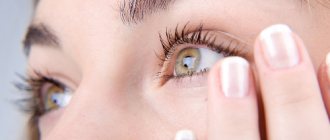
Despite the fact that carp eaters have eyes, they do not perform the same function as in more developed animals. Vision mainly serves to determine the intensity of light, which in turn is related to the temperature of the water. Where it is lighter, it is usually warmer, and carp eaters are warm and light-loving. When attacking a fish, the eyes do not play any role.
Argulez carpied fish photo
Numerous sensitive bristles located on different parts of the body help carp eaters navigate in space, with the help of which they perceive the movement of water and, partly, odors. You can do a simple experiment. Place a strip of paper in an aquarium with carp eaters and move it in the water. The crustaceans rush at the piece of paper, but as soon as they touch it, they swim away. But if you first rub it on the fish, the carp eaters linger longer until they use their mouth proboscis to detect the forgery.
When chasing a fish, carp eaters quickly move in the same direction, parallel to it, and then sit on the head of the victim. Slowly, they crawl to parts of the body that are less washed by water, and settle behind the gill covers and at the pectoral fins. There the covers are relatively thin.
Having attached themselves to the fish, the crustaceans continue to vigorously work with their swimming legs, creating a flow of water necessary for respiration. Carpoeds do not have a heart, but thanks to contractions of the muscles of the abdominal region and intestines, blood constantly circulates in the body cavity.
During the mating season, the male fertilizes the female that has attached itself to the fish, holding her hind thoracic legs with his legs. Then the female leaves the fish and goes in search of underwater plants, stones or other suitable substrate, on which she lays a double row of eggs (from 20 to 300 pieces), gluing them with a special substance.
Depending on the water temperature, after 3-5 weeks, young, but not yet fully formed crustaceans emerge from the eggs. Their swimming legs are underdeveloped, but they have long rear antennae used for swimming. Using the same antennae, as well as the terminal spines of the front jaws, the larvae attach to the fish. They molt twice within a week, and with each molt the rear antennae shorten and the swimming legs develop. From the third to fifth molt, powerful suckers are formed from the front jaws, after which the crustacean enters the sexually mature stage and gives rise to a new generation of parasites.
Argulez carpied fish photo
The full development cycle of carp eaters at a temperature of 10-20°C lasts 70-100 days. In warmer water (21-28°C), crustaceans can produce up to six generations per year, that is, the number of parasites from one fertilized female can already reach 20 billion crustaceans in the fifth generation. In aquarium practice, of course, the development of such a number of parasitic crustaceans is unrealistic - if all the fish were destroyed, they would simply have nothing to eat. Data on the rate of reproduction of the crustacean serve as an indicator of what can happen to an aquarium farm if timely control of the parasite is not started.
Carnivores do not show any preference for fish of certain species and can even attack other aquatic vertebrates - newts, tadpoles, etc.
Having attached itself to the victim, the carp eater pierces the host's skin with its proboscis and sucks blood. To prevent the blood from clotting in the wound, the carp eater injects the secretion of its poisonous gland into it. Hemorrhage occurs at the injection site and an inflammatory process develops. An ulcer forms on the damaged area, through which a secondary infection penetrates.
Argulez carpied fish photo
Subclass copepods (Copepoda)
One of the largest groups of crustaceans, which, according to various estimates, includes from 10 to 20 thousand species. A separate science has even been created to study them - copepodology . Most copepod species are ectoparasites of vertebrate and invertebrate animals, and free-living cyclops and calanoids are the most important component of zooplankton.
Freshwater copepod. Author: Biosciences Imaging Group, University of Southampton/Wellcome Trust Medical Photo Library - Wellcome Collection, CC BY-SA 4.0
Copepods are found in all aquatic habitats, from the deep ocean to the highlands (in meltwater pools on glaciers). Most copepods are inhabitants of the seas, inhabiting the pelagic zone, the seabed and thickets of plants; they form an important link in food chains.
Their role in pelagic food chains is especially important: as small predators, they eat flagellates, diatoms and other unicellular algae as part of phytoplankton, the annual production of which is five times higher than that of terrestrial vegetation, including agricultural plants. Accordingly, the number of some species of copepods is enormous and probably exceeds even the number of common species of terrestrial insects.
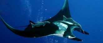
Marine copepods make up the majority of animal protein on Earth; they form the basis of food for giant sharks and baleen whales. Many species of fish, objects of industrial fishing, being larvae, feed mainly on copepods. Some species, such as herring, sprats, sardines and mackerel, feed on copepods even as adults.
Acatocyclops in polarized light. Author: Andrey Savitsky, CC BY-SA 4.0
Producing a huge amount of excrement (one individual can produce up to 200 fecal lumps per day), copepods make a significant contribution to the flow of substances from the water column to bottom sediments, ensuring the prosperity of detritivores. This transfer of substances is especially important for abyssal communities, whose existence depends on “sea snow”. In the marine benthos, among representatives of the meiofauna, copepods occupy the second place after nematodes in number of species and abundance. Here they are a significant food source for flounder and salmon.
In fresh waters, the species diversity of copepods is lower, but here they are just as important. Some representatives of freshwater cyclops (Cyclopoida) are intermediate hosts of human parasites (guinea worm (Dracunculus medinensis), tapeworm (Diphyllobothrium latum)). Other cyclops may be intermediate hosts for fungi and sporozoans that infect mosquitoes and their larvae - such species of copepods could probably be used for biological methods of combating malaria.
Copepods have very different structures and one species cannot be considered as one typical representative. Some parasitic forms are changed so strongly that adult animals could not be classified not only as copepods, but even as crustaceans, if not for the larvae, which leave no doubt about their taxonomic affiliation.
Maxillopods: Cyclops from Lake Linevo, Omsk region. Author: Anastasia Pokhaznikova, CC AT 4.0
Free-living copepods are typically small animals with a body length ranging from 0.5 to 5 mm. Sometimes among planktonic copepods there are species of larger sizes, up to 28 mm long. The largest among the parasitic forms, Kroyeria caseyi, reaches 6.5 cm. They have a teardrop-shaped body consisting of a head, chest and abdomen and large antennae.
Four pairs of their chest legs are flattened and look like oars; they are used for swimming, for which they are called copepods. Some have one pair of vestigial limbs on their abdomen, while others have no limbs. Most have one medium-sized compound eye in the center of the transparent head, usually bright red in color. Subterranean species may be eyeless.
Because of their small size, copepods do not require a heart or a circulatory system (members of the order Calanoids have a heart but no blood vessels), and most also lack gills. Instead, they absorb oxygen on the surface of their bodies. Their excretory system consists of the maxillary glands.
Calanus glacialis (maxillopods). Author: Jasper Nance, CC BY 2.0
The body of free-living copepods consists of a cephalothorax and ten free thoracomeres. The carapace looks only like a cephalothoracic shield. Thoracomeres II–VI usually bear one pair of swimming legs.
The nervous system consists of the suprapharyngeal ganglion, thick peripharyngeal connectives and a weakly dissected chain of ganglia, reaching only to the end of the thorax. The pharynx is short, equipped with strong muscles. The midgut may have a middle (median), unpaired, forward-directed diverticulum (protrusion of the body wall), as well as a pair of lateral diverticula. In many calanoid species, there is a fat sac as a storage of reserve lipids and a means of maintaining buoyancy. The excretory organs are usually nephridia maxilla II. In primitive representatives of the group, the heart is located above the intestine in the pericardium, has three ostia and passes into the aorta, which reaches the head.
Copepods are dioecious. The gonads are paired or unpaired and located on the dorsal side (dorsally). The oviducts and vas deferens open outward on the genital segment. Females have a seminal receptacle connected by a canal with an antrum (bursa) extended inward. The oviduct also opens into the antrum. Males transfer spermatozoa in a spermatophore. Nauplii emerge from the eggs.
Cyclops (Cyclopoida)
An order of copepods, so named for the presence of an unpaired eye in its representatives, a feature similar to that of the mythological hero of Ancient Greece. Distributed in all fresh waters, including groundwater, as well as in the pelagic and benthic zones of seas. They can serve as food for many fish and their young, but they also contribute to human infection with some parasitic worms, primarily the broad tapeworm and guinea worm. They move with the help of swimming legs and single-branched antennules.
Their body is divided into a cephalothorax and abdomen; it does not have a carapace. Cyclops are predators; they feed on rotifers, protozoa, and small crustaceans.
Cyclops (maxillopods), enlarged 50 times. Author: MarekMiš, CC BY 4.0
Drugs for the treatment of argulosis
However, these drugs must be used with caution, since in a therapeutic dose they are detrimental not only to the carp eater, but also to other, often beneficial invertebrates, for example, mollusks, and they are not harmless to the fish themselves.
Chlorophos is added to the aquarium at the rate of 1 g per 100 liters. water for a period of 24 hours, after which all water in the aquarium is replaced with fresh water. During treatment, the aquarium must be shaded.
Karbofos is added at the rate of 0.01 g per 100 liters. water for a period of 24 hours, and then the water is also replaced. This drug cannot be used if the pH of the water in the aquarium is above 8.0 or the temperature is above 30°C.
ADDITIONAL NOTE ABOUT TREATING FISH.
This note outlines brief postulates and thoughts on treating fish for beginners and aquarium hobbyists. Read them carefully, they will help you and your pets.
1. Assess the feasibility of treatment. Sometimes the cost of medicine (500 rubles) is several times higher than the cost of a fish (neon 50 rubles). No matter how cruel it may sound, there is common sense in what has been said. We are only FOR it if you are responsible for those you have tamed and treat the fish as a family member and all that. But no one has canceled the concepts of rationality and expediency.
2. Before any treatment, 1/4-1/2 of the water is replaced with fresh water. This is done in order to minimize the concentrations of nitrogenous compounds - poisons NH3/NH4, NO2, NO3. It is important to understand that often these poisons are the root cause of fish diseases. The fish are poisoned by them, their immunity drops and the pathogenic flora calmly attacks the weakened body.
3. In addition, it is advisable to always have on hand drop tests for the above poisons + PO4 phosphates. For what? Firstly, to know the concentrations and not allow them to be exceeded. Secondly, it is not always necessary to completely zero out nitrate (NO3) and phosphate (PO4), for example, in a planted aquarium, zeroing them out will lead to additional problems with the plants, because Nitrate and phosphate are the main plant nutrients. Thirdly, the tests themselves allow not only to monitor the situation and understand the root cause, but also to control the situation before, during and after treatment.
Remember - you can’t administer medications when nitrogen and phosphate levels are high! By doing so, you will only aggravate the situation, since in addition to the poisons you will also add drugs that both heal and destroy. That is, they have not only positive properties, but also negative ones. Remember that any medicine is not a panacea, not a magic pill.
What tests to use? At your discretion, but preferably in drops and not in strips, because... drip ones are more accurate. In principle, take whatever tests you find, for example, Tetra tests, which are certainly available in every offline store. If these tests are expensive for you, it is quite possible to find our domestic inexpensive tests from VladOx at retail, if time is of the essence or you want to take tests for the future, then our recommendation is UHE tests (sold only online), also domestic, also inexpensive. Each of the above tests has its own specifics, incl. look and think for yourself. The note is not about tests, but about the treatment of fish.
4. How, with what and for what to treat? Any forum discussions about fish disease are fortune telling on coffee grounds. Since an ichthyoptologist can make an accurate diagnosis after scraping the fish and analyzing it under a microscope, or even after dissecting the fish. So, something like this, friends. Our position on the mechanism of home treatment is described in this article, take a look. The bottom line is briefly: if the symptoms of the disease are obvious, as, for example, with ichthyophthyriasis or hexamitosis, then we treat with drugs and according to the treatment regimen for this disease. If the symptoms are not obvious, we treat comprehensively.
Complex medications, for example, Tetra Contralck (contains malachite green and formaldehyde) help well against invasion (parasites - ichthyphothyriasis, oodinosis, costiosis, etc.), but also have a detrimental effect on other pathogenic flora. No, Tetra Contralk, there are other complex medicines.
Ethacridine lactate - 836.0 mg Acriflavine - 160.2 mg Methylene blue - 56.44 mg 9-aminoacridine * HCI * H2O - 28.20 mg
They took them, and, in fact, received the components of Tetra General Tonic. Silva please, gentlemen.
Moreover, having gained experience, you can generally master it and switch to treatment with pharmaceutical drugs.
5. The treatment regimen itself is described in the instructions. There is no special wisdom here. Please note that during treatment it is advisable to increase the aeration of the aquarium and not keep the aquarium lighting constantly on. Many drugs disintegrate quickly when exposed to light.
Of course, there should not be any sorbents (charcoal, zeolite) in the filter; you cannot use water conditioners, such as Tetra AquaSafe or Sera Akutan, they bind drugs.
6. After treatment, do not forget that many medications disrupt the biological balance in the aquarium, that is, they kill both pathogenic and beneficial flora. Here, drop tests, good quality water changes, starter preparations and other measures aimed at restoring the nitrogen cycle in the aquarium will come to your aid.
LET'S SUMMARY. Fish don't just get sick. Negative conditions of detention are the root cause. Either you bought already stunted fish, or negative conditions have developed in your aquarium. Any treatment begins first of all with finding and eliminating the root cause. Next, a decision is made on the treatment mechanism. During the treatment period, special attention is paid to symptoms (regression, progress). Please note that regression of the disease may not occur in 2-3 days. After treatment, we do everything so that the aquarium begins to work as a single, holistic and healthy mechanism.
Argulez video review
[youtube.player]
What is it and how dangerous is it?
Fish louse (carpoeder) is a parasitic gill-tailed crustacean of the genus Argulus. It parasitizes mainly fish.
The fish louse has a special proboscis (stylet), which it inserts into the body of the victim. At the end of the proboscis there is a sharp spike, and on the sides of the spike there are two powerful suction cups that replace jaws. With the help of these devices, the parasite is attached to the body of the fish and is held on it.
The crustacean has 8 legs that can make strong swings. Thanks to this, the crustacean swims quickly.
The parasite has 5 eyes - 2 large, and between them 3 small. But vision does not play a role in finding a victim. The fish louse is guided by the movement of water created by the fish, as well as by smell.
The argulus is attached to the head of the fish, from where it then crawls to other parts of the body where the skin is not very thick. A thick layer of skin does not allow crustaceans to pierce holes in it and suck blood.
They are most often attached behind the gill covers and pectoral fins. Immediately after attachment, the parasite inserts a stylet into the fish’s body, with which it pierces a hole to suck out blood.
At the same time, the poisonous secretion of the parasite enters the fish body, which has a toxic effect. Because of this, the fish loses weight, loses mobility, and its scales become dull. At the puncture site, ulcers and wounds form, through which infection, swelling, and inflammation can penetrate. Most often, crustaceans choose young fish. Argulus affects aquarium fish of all types. If treatment is not started in time, it can lead to mass death of individuals.
Pentastomids, tongue worms, or pentastomids (Pentastomida)
This is a subclass of obligate parasitic maxillopods, nothing indicates that they belong to crustaceans; they do not even have the larvae characteristic of it. But many scientists are inclined to believe that their body and life cycle underwent such a change due to a parasitic lifestyle. More than 100 species of quinques are known, distributed mainly in tropical regions. However, some species are also found in Europe, North America and Arctic regions (bird parasites).
They were called tongue-shaped for the similarity of their body to the tongue of vertebrate animals, pentamouth - for the presence of five front protrusions with appendages, one of them is a mouth, the rest are hooked limbs with which they are attached to the host’s body.
Pentamouths (maxillopods)
Externally and internally they are more similar to annelids:
- worm-shaped body, from 1 to 14 cm long. Females are larger than males;
- skin-muscle bag;
- lack of respiratory and circulatory organs. The constant movement of hemolymph is ensured by peristaltic contractions of the body;
- there are no special excretory organs. Excretion may be through the skin and midgut, or through glands, many of which are located in the skin, on the head, or in the anterior part of the torso;
- Pentamouths have no eyes.
Chemical sense organs and mechanoreceptors are represented by numerous warts. Adult animals of all species are parasitized by:
- in the respiratory organs (lungs);
- respiratory tract (nasal passages);
- adjacent cavities (brain ventricles, air sacs) in carnivorous terrestrial vertebrates (the only exception is still the herbivorous reindeer).
The life cycle of some of them changes hosts. 90% of all species of their hosts are reptiles, the rest are carnivorous mammals (mainly canines) and birds. Development takes place in vertebrates or, in some cases, in insects (cockroach, rhinoceros beetle). A person can also accidentally become infected and become the host of adult dog parasites (Linguatula) or, more often, larvae of various species; they cause linguatulidosis .
Reasons for appearance
Argulus does not start in the aquarium initially, but gets there from the outside:
- Along with food. This is the most common reason for the appearance of this parasite in an aquarium, especially if the food is from natural reservoirs.
- Along with vegetation. Those plants that are planted in an aquarium for the purpose of decoration may harbor parasites that later attack the fish.
- Together with soil or water.
Symptoms
It is not so difficult to notice a crustacean in an aquarium - its size is 3 - 6 mm, so it will be visible to the naked eye. If you still cannot visually detect the crustacean, you should pay attention to the following symptoms:
- Inhibited development of fish, especially young individuals - small size, exhaustion.
- Fish weight loss.
- Ulcers, wounds on the surface of the body.
- Inflammation, secretion of mucus from affected areas.
- The fish becomes inactive.
- Behavior changes - the aquarium resident begins to hide in algae, burrow, and rub against various objects.
How to get rid of a parasite
If a carp eater is found in the body of the inhabitants of the aquarium, it must be removed mechanically:
- Thin tweezers can be treated with any antiseptic.
- Any clean rag, piece of fabric or cotton wool is generously moistened with water - you will need to put the caught fish there.
- The affected fish is carefully caught and placed on a wet surface.
- Using clean tweezers, the louse is removed with utmost care so as not to further damage the body of the affected fish.
- The affected areas are treated with a weak solution of potassium permanganate.
Attention! When working with toxic chemicals, you must follow safety rules and extreme caution! Do not allow substances to come into contact with mucous membranes, skin, eyes, or mouth. When working with them, it is recommended to use protective gloves, goggles and a cotton-gauze bandage (respirator). You cannot breathe in the vapors of these substances! The drugs should be stored in a tightly sealed container, out of reach of children.
The first treatment of fish is carried out in an infected reservoir, then the fish are transferred to another aquarium or any other container and repeated disinfections are carried out there. It is better to keep the fish in another container for a week, and rinse the infected aquarium, dry it and expose it to sunlight. If there are eggs on the glass of the aquarium, then without water all the embryos will die within a day.
If the fish infection occurred not in an aquarium, but in a garden pond, then you need to leave the pond without fish for 1 month. During this period, both old and newly hatched carp eaters will have time to die without an owner.
How to kill salmon sea lice?
Scientists involved in the parasitic fauna of fish consider the most successful method to be the simple destruction of infected fish by burning. This is a very expensive method. Naturally, all fish farms do not budget for such expenses. When the income is only from sales, it will not be possible to take out a loan for the destruction of fish. No commercial bank can afford this. In such a situation, it is easier for farms to leave infested cages alone, hoping that the lice will be destroyed on their own. And then it becomes clear why the salmon sea louse is multiplying more and more in the northern seas.
Many foreign companies immediately detect fish infection when there are only a few such individuals in the cages. This serves as a signal to treat the fish with antibiotics and pesticides; sometimes even fish feeding on Lepeophtheirus salmonis, of the genus Ballan Wrasse, can be added to the cages. Of course, all measures are being taken to catch infected fish, but the parasites multiply at tremendous speed.