Aquarium fish, like any other creature on the planet, are susceptible to various diseases. And one of the most common ailments is hexamitosis - it not only negatively affects the external beauty of the inhabitants of the aquarium, but can also lead to the most tragic consequences in the future. To avoid this, in this article we will tell you not only what hexamitosis (hole disease) is in aquarium fish , but also consider the reasons for its occurrence, symptoms, and effective treatment methods.
Hexamitosis (hole disease) in aquarium fish
Introduction
Without going into details of the cell structure of Diplomonads (which the reader can familiarize themselves with - Poynton, Sterud, 2002), I will note interesting points raised by the authors of this article: - identification of the species Diplomonadina, family Hexamitidae is impossible without studying the ultrastructural features of the cell. Transmission electron microscopy (TEM) is required; — all modern studies using TEM technology have found flagellates of the genus Spironucleus in fish, but not Hexamita. Those. confirmed cases of fish infection only by representatives of the genus Spironucleus.
Schematic illustration of the structure of Diplomonas of the family Hexamitidae. The genera Hexamita and Spironucleus have packets of flagella at their junction, while Octomitus lacks them. The size and shape of the double cell nuclei, the location of kinetosomes at the base of the flagella, and the flagellum exit channel at the back of the cell differ. Small elements are not shown for simplicity. The length of flagella in real Diplomonas is much longer
Diplomonas flagellates prepared using various techniques and viewed under a light microscope.
Drawing. Diplomonas flagellates prepared using various techniques and viewed under a light microscope. (a,b) Fresh specimens of the spherical and elongated trophozoite Spironucleus torosa (Nomarski interference microscope); (c,d) crude preparations of Spironucleus torosa stained with Protargol (colloidal silver preparation, protein silver), marked by a V-shaped structure at the posterior end of the cell [in the TEM image depicted as two rings of microtubules supporting a protrusion near the body, recurrent flagella extending near cell center]; (e) Preparation of Spironucleus torosa stained with Protargol (colloidal silver preparation, protein silver) using the filtration method, highlighting the dark area at the posterior end of the body (the TEM image shows an extensive symmetrical system of microtubules supported by an elongated structure of intersecting ribs at the posterior end of the body) , tubercle, passage of recurrent flagella under the cell surface; (f) unidentified Diplomonas (arrows) in a blood smear stained with Leishman-Giemsa, showing the nucleus and the path of recurrent flagella through the cell; (g) culture of S. barkhanus, stained with 4′-6-diamino-2-phenylindole (DAPI), observed under light field and UV radiation, the nucleus appears blue-violet (white in the photograph); (h) Spironucleus torosa in tissue sections (rectal cavity), stained according to Feulgen (Feulgen reaction); (i) Spironucleus torosa in tissue sections (rectal cavity) stained with hematoxylin-eosin. Abbreviations: (cp) caudal projection; (p) tubercle; v – V-shaped structure at the posterior end of the body (scale = 10 µm).
Habitat
Single-celled flagellates can live in the environment, especially contaminated water and sediment, or be parasites of invertebrates (molluscs, crustaceans), fish, amphibians, birds, mice and other mammals. Their main function in their environment is to digest bacteria to obtain nutrients.
The most famous diplomonas are Giardia, which is the most common cause of diarrhea in people around the world. This organism can also cause death in young children or immunocompromised people. The parasite attaches to the upper wall of the intestine and, multiplying, begins to be excreted in the feces. It is resistant to disinfectants such as chlorine in swimming pools. Cysts remain in water for a long time, and ingestion of even a small number of them leads to infection. In fish, infections with another flagellate, Spironucleus, occur in a similar manner. It infects Arctic char, trout, wild and farmed fish, other tropical aquarium species, mice and birds.
Successful control of infection depends on understanding the biochemical differences between the pathogen and its host, in particular the formation of the cyst wall. Diplomonas prefer an environment with low oxygen content; they are poorly protected from exposure to air. Thus, the introduction of oxidizing agents (ozone, active forms of chlorine, peroxides) into water will help fight them [Lloyd, Williams, 2014].
Treatment
Today, there are several options for ridding fish of this disease. But it is worth emphasizing that it is necessary to choose which method to use based on what became the catalyst for the development of the disease. Thus, it has been scientifically proven that hexamitosis is almost always accompanied by a viral infection. Therefore, remember that thoughtlessly started treatment with metronidazole can lead to the most unexpected consequences. Let's look at how this disease is treated.
First of all, you need to move the infected fish from a common artificial reservoir to a separate vessel, which will serve as a kind of quarantine. This action is necessary in order to avoid the development of the disease throughout the aquarium. After this, it is recommended to slightly increase the temperature of the aquatic environment in the fish tank. Ideal temperature values would be 34-35 degrees.
Such a sharp jump can have a detrimental effect on some parasites and cause their death. But you should be careful and before performing such an action you need to familiarize yourself with the physiological characteristics of your pets, because not every fish can be suitable for high water temperatures. For example, treating cichlids this way will not bring any results.
Another option for ridding fish of the manifestation of this disease is treatment with metronidazole. This antiprotozoal drug has already proven its effectiveness. Also, due to the fact that it contains substances that do not affect the environment, it is not at all surprising that many aquarists use metronidazole.
It can be used both in a general artificial reservoir and in a quarantine pond. But it is worth emphasizing that the maximum dose of the drug should not exceed 250 mg/35 l. It is better to use metronidazole for 3 days, while performing regular water changes in the ratio of 25% of the total volume on 1 day, and 15% on subsequent days. If the treatment does not bring a noticeable effect, then it is more advisable to suspend it.
The first results of taking this medicine will be noticeable after the first week. Also, for prevention purposes, it is advisable to repeat the treated baths after 1 week.
In addition, in addition to metronidazole, you can use other special medications, which can be purchased at any pet store. But before you make a purchase, it would be useful to consult with the seller whether their use will harm the established microclimate in an artificial reservoir.
So, among the most popular are:
- tetra medica hexaex;
- zmf hexa-ex;
- Ichthyovite Kormaktiv.
It is also worth noting that the greatest effect in the fight against this disease can only be achieved with an integrated approach.
So, as mentioned above, some fish can only be carriers of the pathogen, unlike others. Therefore, you should not treat fish with only one drug. But even here you should be careful. Thus, experienced aquarists recommend treating hexamitosis using both pharmaceutical and branded drugs. For example, 50 mg of Furazolidone should be used per 15 l, simultaneously with the drug Kanamycin (1 g/35 l). Use every day for a week with regular replacement of 25% of the total water volume.
If the drug Ciprofloxacin is used, its dose is calculated in a ratio of 500 mg/50 l. It is best to use ZMF HEXA-ex at the same time. How to dilute this drug can be found by reading the instructions.
Sometimes, after treatment, some fish may show signs of toxicosis. In this case, it is necessary to urgently replace at least half of the water in the artificial reservoir and subsequently use half the dose of medications. This requirement applies to both branded products and those purchased at the pharmacy. [important]Important! Upon return of quarantined fish, it is recommended to carry out preventive measures in a common tank over the next 4 days to avoid a possible relapse.
Infection and spread of hexamitosis
The causative agent of hexamitosis infects wild and cultivated fish, including aquarium cichlids (discus, angelfish, astronotus). The parasite settles at the beginning of the intestines, immediately after the stomach.
Most likely, flagellates are transported horizontally. Cysts or trophozoites (vegetative stage of development) enter the body with water contaminated with feces. Flagella allow the parasite to reach the upper intestine. In the intestinal tract, diplomonas move freely in the lumen and feces.
The flagellate generation time is 24 hours. With the blood, the parasite penetrates other parts of the body, and its population increases.
In addition to infection of internal organs, infection of the body, the appearance of ulcers and erosion of the lateral line cannot be ruled out.
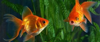
Among freshwater aquarium fish, Perciformes (cichlids) are especially susceptible to the causative agent of hexamitosis. Carps - koi and goldfish - get sick less often.
It is worth noting that normally the causative agent of hexamitosis is often found in small quantities in fish. Based on available data, it can be assumed that S. vortens, the recognized causative agent of cichlid hexamitosis, is intermediate between a commensal organism and an opportunistic parasite. For example, healthy angelfish often have this pathogen in their intestines, but systemic infection does not develop. Moreover, ulcers with hole disease often contain bacteria. Those. Flagellates may indirectly accumulate in ulcers, feeding on these bacteria, rather than causing the lesions themselves [www.biomedcentral.com/1471-2180/8/71].
Prevention measures
As mentioned above, hexamitosis develops when optimal conditions for it appear in an artificial reservoir. Therefore, preventive measures consist of constantly maintaining an ideal ecological balance in your artificial reservoir.
In addition, it is recommended to periodically feed the fish with certain medicinal foods containing substances such as spirulina, kanamycin and furazolidone. Also, you should not constantly use the same type of food. It would not be superfluous to purchase Fishtamin or Activant preparations for an artificial reservoir and then add them to the aquatic environment.
Prevention of hexamitosis
On a note ! You should also be very careful not to overfeed your pets and do not forget to check the level of nitrates in the aquatic environment.
Remember: hexamitosis causes almost irreparable damage to the digestive system of fish, which can ultimately lead to death. Therefore, following these simple recommendations can not only save the life and health of all inhabitants in an artificial reservoir, but also save you from unnecessary expenses on expensive medications.
Symptoms of hexamitosis
Weak and stressed individuals are most susceptible to infection. With hexamitosis, fish lose appetite and weight. She hides in secluded places and stops moving normally. The color darkens, the head becomes lighter than the rest of the body. Often, due to emaciation, the head of the fish appears larger than its body (the so-called “pinhead” fish).
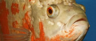
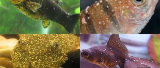
External manifestations of hexamitosis in astronotus (Astronotus ocellatus) (mishka, www.oscarfishlover.com/forum)
Manifestations of hexamitosis in the Flowern Horn cichlid (Alamy, practicalfishkeeping.co.uk)
External manifestations of the disease are called “holey disease”. A whitish mucus is secreted from the gills, mouth, ulcers, head and anus of the fish. After 48 hours, the eyes become coated with a whitish coating. After a few days, individuals with cataracts lose their vision. At the final stage of hexamitosis, fish are faced with a lack of oxygen; their gills are filled with blood so that the transfer of oxygen to other parts of the body is difficult. Individuals swim to the surface of the water and then return to the bottom. Movements are uncoordinated.
After 14-16 days, in the absence of treatment, patients usually die. At all stages of hexamitosis, infected individuals release parasites into the water and spread the infection. For this reason, fish farmers need to wash their hands after working with sick people.
A chronic course of hexamitosis is possible. It is often observed in cultivated fish in spring and autumn. The mortality rate among them is slightly higher than that of healthy fish, however, due to the long cultivation period, losses can be significant [Tojo, Santamarina, 1998].
Causes of the disease
The most common reason for the active spread of diplomonas, leading to an outbreak of hexamitosis in fish, is pollution of the reservoir.
Factors that provoke hexamitosis in fish raised in ponds:
- decrease in oxygen concentration in water;
- eating disorder;
- monotonous feeding or sudden change in diet;
- a lot of fish per unit volume of reservoir;
- keeping fish of different sizes in one pond;
- excess nitrates in water;
- vitamin deficiency due to deficiency of vitamins, especially groups B and C;
- stress caused by certain manipulations, for example, moving from pond to pond, transportation.
Hexamitosis most often affects salmon fish. In them, the disease usually manifests itself on the sides and scalp. Carp and grass carp are also susceptible to infestations. Catfish and eels can become victims of these parasites.
Diagnostics
Accurate diagnosis of hexamitosis is carried out through microscopic examination of crushed intestinal preparations (200-400x magnification) or feces. Flagellates move quickly and erratically. They are easy to notice in the area of mucosal detachment and in cases of serious infection [Sahandi, Hajimoradloo, 2011].
In living individuals, after anesthesia, light pressure on the abdomen squeezes out the feces from the intestines. The sample is placed on a glass slide and three drops of water are added. Cover with a coverslip and examine under a microscope (400x).
The degree of parasite invasion can be assessed using the following scale [Tojo, Santamarina, 1998]: Zero (-) - there is no causative agent of hexamitosis on a glass area of 24x32 mm; Minimum (+/-) - only one parasite was detected on a glass area of 24x32 mm; Low (+) - more than one parasite was found in the entire specimen (24x32 mm), less than 10 individuals in the microscopy field; Moderate (++) – 10-50 individuals in the microscopy field; High (+++) - >50 individuals in the microscopy field
Autopsy
From dead fish, the intestinal area immediately after the stomach is removed. In sick individuals, this section has a bright yellow color (due to the secretion of bile). Pressed intestinal preparations are prepared and examined under a microscope.
The fins of the fish are dark and have hemorrhages at the base. The scales are covered with whitish, yellowish mucus. The intestines, liver, spleen, swim bladder, heart, skin and fins are affected. However, histological changes in the internal organs are observed only in some species. Thus, in angelfish, granulomas form in the liver, and infiltration of macrophages in the spleen and lymphocytes in the intestine occurs. This is not observed in Astronotus and Mbun.
Before death, numerous internal hemorrhages occur.
Tissue from the liver (1), spleen (2) and intestine (3) of an angelfish infected with hexamitosis. Hematoxylin and eosin staining. Scale 50 and 100 µm. Gr - granuloma, M - macrophages, * - area of inflammation, lymphocyte infiltrate and submucosa in the intestine (Suchanya Mankhakhet et al., 2012)
To identify the causative agent of hexamitosis during intestinal autopsy, it is recommended to first leave the fish without food.
It is worth noting that fish severely affected by the causative agent of hexamitosis (numerous parasites in the pyloric pylorus of the stomach and intestines) may still lack damage to the mucous membrane of the skin and external signs of invasion into the epithelium [Tojo, Santamarina, 1998].
Symptoms
As mentioned above, it is almost impossible to recognize this disease at the initial stage. That is why it is quite problematic to start timely treatment. The only indirect signs can be considered a darkening of the fish’s natural color, sudden loneliness or weight loss, despite the fact that it eats regularly. If such signs appear on the face, then experts recommend immediately examining your pet for the development of an unwanted disease, so that the subsequent treatment provided is effective.
Also, in addition to this, we will consider the main symptoms of the development of this disease in a common aquarium. This is how they are classified:
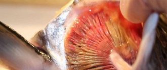
redness of the lips is a sign of hexamitosis in fish
- Decreased appetite. In a more acute form, even a complete refusal to eat food is possible.
- Pickiness when eating. So, the fish may first grab the food, but then spit it out.
- The appearance of white mucous discharge. This occurs due to the fact that the disease affects the pet’s intestines, which leads to the rejection of its cells, which are released in large quantities from the fish’s body. Also sometimes, hexamitosis can cause indigestion. Because of this, you can observe a picture where undigested food is also excreted along with waste products.
- Abdominal bloating. But, as a rule, such symptoms can be observed mainly in cichlids. Most often, this disease causes changes in the shape of the belly and back of fish.
- The appearance of deep erosions on the lateral areas of the fish, extending to the skin of the head.
- Anal enlargement.
- Destruction and loss of fins.
And this is not to mention the changes that the external color of the inhabitants of an artificial reservoir undergoes.
It is worth noting that in most cases, hexamitosis is not characterized by all of the above volumes. Sometimes, white discharge may even indicate the development of enteritis or poisoning. But it is also not recommended to neglect what you see. The ideal option would be to move the infected pet into a separate vessel for examination. In this case, not only is the ecological microclimate in the aquarium not disturbed, but there is also a high probability that the treatment with metronidazole will be effective.
Hexamitosis in cichlids and other fish species
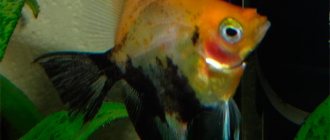
The photo shows hexamitosis in an angelfish (Pterophyllum scalare) (advancedaquariumconcepts.com/hole-in-the-head-discus-oscars.html)
Reliable results of infection of angelfish, astronotus and mbuna with the species Spironucleus vortens were obtained. It was possible to obtain a culture of the parasite from an angelfish. In a fresh preparation, it was active and quickly rotated around the longitudinal axis. Trophozoites were pear-shaped, oval, 9.0-16.0 µm (average 12.86 µm) in length and 3.0-10.0 µm (average 6.52 µm) in width. They carried an S-shaped nucleus and 8 flagella (6 anterior and 2 posterior).
Artificial infection of angelfish with hexamitosis led to the death of 13.33-86.67% of individuals within 14 days. The clinical manifestations of the disease were similar to those of wild specimens, but some aquarium specimens died without any clinical symptoms.
In experiments with artificial infection of guppies, swordtails, goldfish and platies, resistance of these species to Spironucleus vortens was demonstrated. The causative agent of hexamitosis has not been detected in cockerels (Betta splenders).
In another study, it was shown that hexamitosis of severums, angelfish, and Nile tilapia is also caused by flagellates of the species Spironucleus vortens. The parasite was isolated from the kidneys, liver, spleen and head ulcers of fish [Paull, Matthews, 2001].
On the other hand, cold-water cultured fish, Atlantic salmon, Arctic char suffer from the species Spironucleus barkhanus [Guo, Woo, 2004], and rainbow trout from the species Spironucleus salmonis. The course of hexamitosis in them has two phases - blood and tissue.
Thus, the causative agents of hexamitosis are numerous and demonstrate clear species specificity to the hosts.