The causative agent of costia (ichthyobodosis) is Costia (Ichthyobodo) necatrix , a single-celled flagellate parasite. Here is what is written about these parasites in the fundamental guide to zoology published by the Russian Academy of Sciences: “Bdonids, the reticular mitochondria of which contain several kinetoplasts. Ectoparasites of fish, attached to the host epithelium by a modified anterior end of the body (rostrum)"
.
Names of costiosis in foreign literature: Costiosis, or Ichthyobodosis.
Symptoms
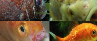
The affected fish first begins to rub against various objects, then refuses to feed. The introduction of the parasite causes severe irritation of the integument and the release of mucus. Dull bluish-grayish spots appear on the body of sick fish, which subsequently merge into a solid gray coating due to abundant mucus secretion. The most infected areas of the skin, where parasitic fungi settle, undergo decomposition. The gills turn pale and become covered with mucus, as a result of which breathing and gas exchange are disrupted, and the fish rise upward and swallow air. The fins are glued together. Pisces react poorly to external stimuli and become restless. In the absence of timely treatment, massive fish death may occur.
Photo: Costia or Ichthyobodo
Molluscum contagiosum in children
Molluscum contagiosum is more common in children than in adults. Children up to one year old practically do not get sick with molluscum contagiosum. This is due to the fact that in the first year of life the child’s circle of contacts is small: the child moves in a limited space, often specially prepared for him and under the strict supervision of adults, without trying to come into contact with other children. But as soon as a child begins to actively communicate and independently explore the world, the threat of becoming infected with molluscum contagiosum increases sharply.
The peak incidence of molluscum contagiosum in children occurs between the ages of 2 and 6 years. Immunity at this age is still weak. Children become infected through toys or dirty hands. The virus penetrates the skin in the place where the integrity of the skin is damaged - through wounds, abrasions, cracks. Children's skin is delicate and sensitive, and the activity of a preschool child is great. As a result, numerous microtraumas occur, opening the way for infection. Cases of infection with molluscum contagiosum while swimming in the pool have also been described.
From 6 to 10 years, the incidence of molluscum contagiosum decreases. Instilling household hygiene skills is of great importance. The sooner your child starts taking care of clean hands, the better.
Causes
Invasive disease. The causative agent is the flagellate Costia necatris, the most dangerous ectoparasite that parasitizes the skin and gills. The body shape is pear-shaped, length 0.01-0.012 mm, width 0.006-0.008 mm. It is attached to the skin of the fish with the help of two flagella, which are used for swimming. Reproduces by division. It enters the aquarium from a reservoir where fish are found along with food, plants and soil, if it has not been boiled or calcined, as well as with fish, shellfish, plants, water and equipment from an infected aquarium. Mass development of the flagellate occurs at a temperature of 20-30 degrees. When unfavorable conditions occur, it forms dormant forms and cysts that are resistant to influence and are a source of possible infection. The disease is most dangerous for fry. Poorly nourished fish tolerate the disease with difficulty, especially in acidic, poorly aerated and irreplaceable water. Well-fed fry and adult fish tolerate costiosis relatively easily.
Costiosis of fish
A dangerous disease of juvenile farmed fish, widespread in the southern and central fish farming zones.
Pathogen . The causative agent is the flagellate Costia (= Ichthyobodo) necatrix from the family. Bodonidae.
The causative agent of fish costiasis is Costia necatrix.
This is a small oval with a slight concavity (or wedge-shaped - from the side) parasite, 5 to 20 microns long, with two long elastic flagella with which it moves. The parasite attaches to the host using finger-like projections formed at the anterior end of the body, with which it penetrates the cell and sucks out its contents. Localized on the surface of the body and gills. Costia feeds on bacteria that appear on the dying skin tissue of fish, dying and living cells of epithelial tissue. The parasite reproduces by longitudinal division. Withstands temperatures from 2 to 30 ° C, the optimal development temperature is 25-26 ° C. When unfavorable conditions occur, the parasite forms cysts. The reproduction of flagellates is facilitated by the acidic reaction of water.
Epizootology . The disease affects juvenile carp, salmon and other fish in fish farms in various climatic zones of the USSR. Fish of older age groups rarely get sick and are parasite carriers. Most often, the disease occurs in spawning ponds or during the rearing of larvae in trays and apparatus at high stocking densities, insufficient nutrition, and starvation. In nursery ponds, where the stocking density is lower and the food supply is better, the disease subsides. In recent years, bone disease has been observed in wintering fish in pools and ponds at temperatures of 0.5-3°C. Sometimes costiosis is recorded together with chylodonellosis, which was noted in one of the fish farms of the Moscow region in March-April in white and bighead carp and their hybrids. Low pH values, which occur in reservoirs on peaty soils, are favorable for the development of the disease. Parasites enter fish ponds with water and are introduced by wild fish from water supplies. Uncontrolled transportation of fish contributes to the spread of the disease.
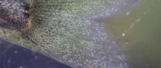
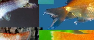
Clinical signs . Sick fry come to the surface of the water and accumulate on the water supply. A bluish-matte mucous coating appears on the body of the fish. As the disease progresses, the fish lose weight, become inactive, do not respond to external stimuli and are carried by the current of water to the water outlet. Their gills are covered with a thick layer of mucus, pale, sometimes necrotic. With severe damage, the interfin membrane is destroyed and the fin rays are exposed.
Diagnosis . The diagnosis is made after microscopic examination (under high magnification) of scrapings from the surface of the body, gills and detection of a large number of parasites.
Control measures . To prevent disease, juvenile fish must be provided with a sufficient amount of natural food. Excessively dense planting and overexposure of fish in spawning and fry ponds should not be allowed. Juveniles are transplanted into nursery ponds as early as possible, creating a good food supply there. Before filling, spawning ponds must be treated with quicklime (at the rate of 25 c/ha) or chlorine lime (at the rate of 5 c/ha). Producers should be treated 2 weeks before planting for spawning in a 5% solution of table salt (5 min) 3 times every 5-6 days.
Good results in the treatment of trout fry are obtained by treatment in baths: brilliant green at a dilution of 1: 200,000 for 10-15 minutes; table salt with a concentration of 2.5-3% for 3-5 minutes; formalin at a dilution of 1:4000-20,000 for 10-15 minutes, quicklime and potassium permanganate with a concentration of 5 g/m3 for 20 minutes. In baths of malachite green with a concentration of 0.1 g/m3, costia dies within 24 hours.
If you find an error, please select a piece of text and press Ctrl+Enter.
Treatment
The fish are transplanted into a quarantine aquarium, in which they are treated with malachite green, or mytelene blue. And in a general aquarium, especially when the water temperature rises to 32-34 degrees, flagellates die within 24 hours. Fish from a quarantine aquarium can be treated in separate baths: 0.1 g of potassium permanganate per 10 liters of water, hold for 40-50 minutes; 1 g of copper sulfate per 10 liters of water, exposure 10-20 minutes; 150-200 g of table salt per 10 liters of water, soak for 10-15 minutes. Note: When treating with medications, the water temperature should be at 24-25 degrees. When treating with table salt, it should be taken into account that not all fish tolerate such a high concentration.
Methods for diagnosing molluscum contagiosum
Molluscum contagiosum can be confused with manifestations of other diseases, including serious ones such as syphilis or cancer. Also, the activity of the molluscum contagiosum virus increases with a decrease in immunity, so in 20% of cases molluscum contagiosum accompanies HIV infection. This means that when rashes appear that correspond to the description of molluscum contagiosum, a medical examination is required to rule out such options.
When contacting a dermatologist, the doctor will examine the patient, make a diagnosis and suggest a treatment method.
Inspection
In most cases, the diagnosis of molluscum contagiosum is made by a dermatologist based on the results of an examination of the patient.
PCR diagnostics
Since HIV often accompanies molluscum contagiosum, PCR diagnostics for HIV can be prescribed.
More information about the diagnostic method
Serological blood test
When molluscum contagiosum is detected in adults, a serological blood test is prescribed to identify sexually transmitted infections (hepatitis B and C, HIV, syphilis, etc.).
More information about the diagnostic method
Sign up for diagnostics To accurately diagnose the disease, make an appointment with specialists from the Family Doctor network.
Dropsy
It is caused by a viral or bacterial infection. This disease is dangerous for fish with reduced immunity.
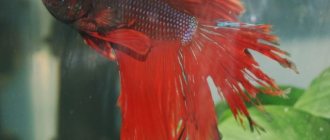
External signs of the disease: the cockerel’s belly is swollen, its scales are ruffled, its belly turns red, and bruises appear on its body. Treatment is prescribed with antibacterial drugs and antibiotics, but the measures are effective only at the initial stage of the disease.
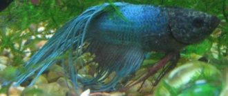
Exophthalmia
This condition is also known as bulging eyes. It can affect either one organ of vision or both at once. Symptoms:
- The eye swells and comes out of its orbit;
- Clouding of the cornea is observed.
To carry out treatment, the fish are placed in a special sterile aquarium and Epsom salts (sold in a pet store) are added to the water. Therapy is carried out until the eye is completely healed.
What to do to prevent your fish from getting sick
Betta fish diseases are much easier to prevent than to treat. Individuals with weakened immune systems and living in an aquarium with unsuitable conditions are more likely to suffer. By following a minimum of rules, you can protect your pets from infection and death:
- Carry out regular water changes, remove food debris from the soil and monitor the condition of the filter.
- Feed the fish only high-quality food and avoid overeating.
- Newly acquired fish and plants must be kept in quarantine for several weeks. For prevention, you can add a little table salt to their water.
- Diseases such as tuberculosis can be transmitted through live food. Therefore, all live food must first be disinfected.
Oodiniosis (velvet disease)
Carried by flagellated parasites. Has characteristic features:
- The appearance of white nodules on the body that resemble dust or flour;
- Ruffled scales;
- Tail peeling.
Also, with oodiniosis, the behavior of the fish changes: it becomes apathetic, lies on the bottom, does not eat and breathes heavily.
To carry out therapy, the sick individual is placed in a separate reservoir. But, since the disease is carried by a parasite, other fish in the general aquarium must also undergo prophylaxis to avoid infection. The following drugs are used:
- Bicillin – 5;
- Copper sulfate;
- Malachite green;
- Formalin;
- Purple K.
The dosage for each drug is described in the instructions and is individual. It depends on the purpose of use - treatment or prevention.