Immediately read about how to treat fish for trichodinosis >>>
Trichodinosis (trichodinosis) is a fish disease caused by round-ciliated ciliates from the family. Trichodinidae - trichodinids (class Oligohymenophora, subclass Peritricha). The name of the disease itself comes from the name of the family, therefore, despite the fact that trichodinids are represented by several genera (Trichodina, Tripartiella, Trichodinella, Paratrichodina, Dipartiella, etc.), fish diseases caused by representatives of these genera are called trichodinosis. Therefore, the widely used name “trichodinosis” cannot be considered correct. However, in foreign scientific literature, trichodinosis also appears under two names: both Trichodiniosis and Trichodinosis are used.
| Videos 1 and 2. The video above shows a typical picture observed when microscopying scrapings from the body of a fish with trichodinosis at low microscope magnification (10*10). Fast moving fluttering “limpets” are trichodines. Below - trichodins at high magnification 40*10. Numerous cilia are visible; it is they that ensure the rapid movement of the ciliate in the water when swimming freely and movement along the surface of the body of the host fish. |
SYMPTOMS OF TRICHODINIOSIS
The symptoms
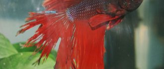
of trichodinosis (trichodinosis) and chylodonellosis are largely similar, so without a microscope it is quite difficult to distinguish trichodinosis from chylodonellosis. In both cases, “foggy” whitish areas are visible on the body of the fish, which arise due to increased secretion of mucus on the affected areas of the skin. Perhaps these areas are more noticeable with trichodinosis than with chylodonellosis, and with very severe invasion, the plaque on the body may be slightly grainy. These “grains” are very small and are noticeable only at 2x or more magnification. They are much smaller than the pustules observed with ichthyophthyriosis and represent both the ciliates themselves and lumps of mucus. On the fins of fish, the trichodins themselves are more visible and high-quality macro photography is enough to see them.
| Photo 1. Photo from the Database of Parasites in Fish and Shellfish Severe trichodinosis in salmon fry. Trichodines are visible on the caudal fin and body. Magnified approximately 25 times. |
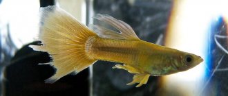
| Photo 2. Trichodinosis in cardinal fish (Tanichthys albonubes), trichodins are visible as very small light grains on the body and fins of the fish. An increase of approximately 5 times. |
| Rice. 1. Trichodins on the respiratory folds of the gill filament of pike. DS - respiratory folds, T - trichodyne. From: Dogel V.A. Parasitic diseases of fish. M.; L., 1932, 149 p. In some cases, trichodins settle only on the gills, then there are no heavily mucus-covered matte areas on the body of the fish. |
When fish become ill, they lose their appetite and mobility. They may begin to itch on the bottom, plant leaves and protruding elements of decoration. Viviparous fish characteristically “sway” from side to side, while standing in one place. Juvenile carp fish crowd together in secluded corners of the aquarium, clinging to the bottom, and if the gills are damaged, they suffocate.
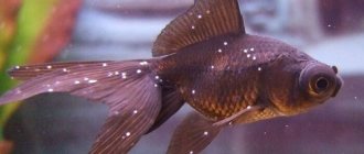
Photo 3. A young cardinal suffering from chronic trichodinosis. Without magnification (the small fish in the inset at a screen resolution of 1366×768 is presented in natural size), the symptoms of the disease are almost invisible. Only the fins have stuck together a little and something can be seen on them. However, the fish do not eat well and die little by little (one per week). But if you enlarge the picture, then the frayed ends of the fins are visible quite clearly, and, most importantly, the noticeable dullness of the color on the body and the graininess on the fins are striking. Unlike chylodonellosis, matte mucus in trichodinosis is noticeable when looking directly at the fish standing sideways in front of the observer. With trichodinosis, in order to see this mucus on the fish’s body, it is much easier to select suitable lighting than with chylodonellosis.
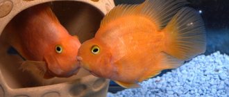
| Photo 4. Since everything is learned by comparison, we present a photo of a healthy cardinal. His fatness and bright shine immediately catches your eye. The fins are intact at the edges, well spread and brightly colored. Two longitudinal bright, rather than dull, colored stripes on the side are clearly visible. It was very difficult to photograph this fish. She is extremely mobile and constantly jumped out of focus. In the end, it turned out far from perfect, but everything you need is visible. But, in general, the cardinal, when he is in good shape, is very handsome, don’t you think? |
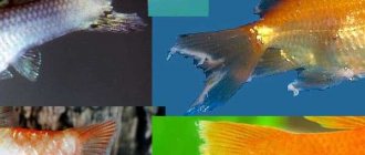
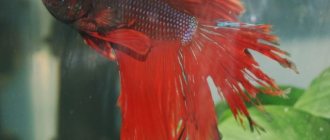
| Photos 5, 6. Symptoms of trichodinosis: stunted growth, exhaustion of fish, destruction of fins along the edges (arrow in photo 5), increased secretion of mucus. Photo 5 - fish in reflected light, photo 6 - in transmitted light. In transmitted light, the symptoms of trichodinosis are more visible. The arrows in photo 6 indicate clusters of trichodynes. An increase of approximately 7 times. | |
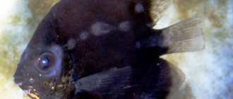
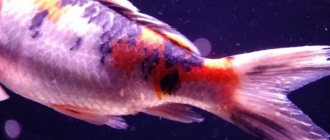
| Photo 7. Young koi carp with acute trichodinosis. Its entire body is covered in trichodynes, an increase of approximately 20 times. The inset shows a life-size fry. |
It should be noted that trichodinosis never occurs for no reason, only because the causative agent of the disease has entered the aquarium. These parasites cannot take root on strong and healthy fish living in optimal conditions. Fish can probably be carriers of individual specimens of parasites, which, due to their small numbers, cannot cause them noticeable harm. It’s another matter if the fish are weakened in some way, then the number of trichodins begins to increase rapidly, they irritate the skin and gills, causing them to become severely slimy. Water pollution with organic substances, for example, remnants of flake food (and high oxidation of water), chronic poisoning with ammonia and nitrites, high crowding of fish, chronic lack of oxygen - these are the factors contributing to the mass reproduction of trichodins. Trichodins do not form resting cysts and are not very tenacious themselves (they can live no more than two days in free swimming without a host fish), so dealing with them is quite simple. It is very important to identify and eliminate the causes of weakening fish. The most likely reason is the unfavorable hydrochemical parameters of aquarium water, especially its high oxidability (see “5 parameters ...”, Oxidability of aquarium water: a simple and very useful test).
Video 3. Microscopy of a temporary wet preparation of a scraping from the body of a young discus patient with trichodinosis - trichodina at low and high magnification of the microscope.
Where does Trichomonas come from?
At the end of the fifties of the last century, several international conferences were held to discuss where Trichomonas comes from in the reproductive tract. They decided that this single-celled organism can be obtained not only through sexual contact, but also from a bath bench or basin, and even from swimming pool water and just tap water, not to mention wells. Bath accessories were blamed because trichomoniasis was practically not detected in men due to the absence of clinical symptoms. If a woman complains of inflammation, and a man is fine, then apparently the woman “caught the infection” somewhere else.
Where can a decent and faithful woman become infected with a sexually transmitted infection? Of course, in a public bath, where she is completely defenseless. And at that time, personal bathrooms were very rare; citizens went to the baths to wash themselves. Another five-year-old mankind was convinced that Trichomonas brought into a family was not at all a consequence of the infidelity of one of the spouses, but evidence of a woman’s failure to observe personal hygiene. One good thing is that this misconception helped save the family, because the infection happened through “non-sexual” means.
But in the mid-60s, scientific proof of the extremely poor survival of Trichomonas outside the human body came from Soviet Estonia, therefore infection by any other method other than sexual intercourse is simply unrealistic. Trichomonas does not like sunlight and hot water, and is immediately killed by any antiseptic. Indeed, Trichomonas is so delicate, so demanding of the external environment that even in the secretion of the genital tract collected for the purpose of research, it often dies before it is brought to the microscope. Since that time, the disease caused by Trichomonas vaginalis has been recognized as a venereal disease.
The main route of transmission of Trichomonas in adults, of course, is sexual, but oral sex does not contribute to infection, in any case, they could not prove this possibility of transmission. A woman in labor with trichomoniasis can infect her child, more often this happens to girls. This route of transmission of infection is called vertical. And today, 2–17% of newborns have trichomonas vaginitis or, in the old way, colpitis inherited from their mother. It is quite possible, because tests are taken from pregnant women several weeks before giving birth.
At risk for trichomoniasis are citizens who have multiple sexual partners, workers in the illegal sex industry, drug addicts and HIV-infected people, socially disadvantaged people, in general, comrades. True, citizens are still actively using the idea of contracting trichomoniasis in the pool, the main thing is to get treatment, and let them decide for themselves how to explain the appearance of Trichomonas vaginalis to their partner.
Formalin (37% formaldehyde)
Formalin is used at a concentration of 15-25 mg/l for the treatment of non-salmon fish. Short-term one-hour baths with a concentration of 125-250 mg/l are allowed. Do not use more than 167 mg/l formaldehyde at temperatures above 21°C. Treatment should be stopped as soon as the fish begins to show signs of stress. Trout is kept for an hour in a solution of 167 mg/l at a temperature of 10-18°C, or more than 250 mg/l at a temperature below 10°C.
Immediately before processing, it is recommended to keep the fish in a solution of table salt in order to increase the separation of epithelial mucus. This mucus acts as a protective barrier for parasites, so reducing its amount improves the therapeutic effect of the drugs. —— www.aces.edu/dept/fisheries/aquaculture/pdf/4701fs.pdf
Ambiphrya and Apiosoma
Ambiphrya and Apiosoma are ciliated ciliates. As adults, the parasites are immobile. They attach to the gills and skin of fish. Ambiphrya (formally, Scyphidia) is barrel-shaped and attached to the integument of fishes by means of many flat scopulae or retaining organs at the posterior end of the body. Around the mouth and in the middle of the body (ciliated girdle) the ciliate has a ring of cilia. Typically, the core of Ambiphrya is ribbon-shaped.
Massive infection of tilapia fry with Ambiphrya ameiuri (top left), distribution of the parasite on the skin (top right) and gill filaments of tilapia (www.sci.sdsu.edu/salton/Ambiphryaameiuri.html)
Apiosoma (formally Glossatella) has a long body with a small attachment site. It does not have a ciliated band, but only an oral ring of cilia. The cell nucleus is more compact, conical or triangular.
These parasites do not damage fish tissue, but in high densities they prevent the supply of oxygen. Like most protozoa, Ambiphrya and Apiosoma are more dangerous in conditions of high organic load and fish stocking density. Ciliates are usually gotten rid of by a single treatment of water with formaldehyde, CuSo4, KMnO4. They may also resolve themselves as environmental conditions improve.
Proliferative kidney disease (Tetracapsula bryosalmonae; formally called PKX)
Tetracapsula bryosalmonae are myxosporidian parasites that cause proliferative kidney disease (PKD) in several species of trout and salmon in western North America and Europe. PKD typically occurs in the spring and mortality is highest at 15°C.
Clinical symptoms include inflammation of the kidneys and spleen, blanching of the gills, darkening of the body, ascites, and bulging eyes. Enlarged kidneys are sometimes noticeable to the naked eye before opening. Inflamed kidneys have a nodular or thread-like surface. This overreaction of the kidneys to infection and the inability of the parasite to form spores in fish suggests that trout and salmon may be unnatural or aberrant hosts of Tetracapsula bryosalmonae. Microscopically, stained tissue smears show the primary cells of the parasite with secondary or daughter cells located within or near the primary cells.
To prevent PKD, fingerling trout should not be stocked until summer. Once infection has developed, mortality can be reduced by increasing the salinity from 0.008 to 0.012 (about 1/3 of the salinity of seawater).
Diagnostics
The presence of Trichomonas in the genital tract must be proven by detection under a microscope or by growing it in a special nutrient medium.
They specifically look for Trichomonas vaginalis in women who complain of discharge from the genital tract, with long-term and drug-resistant inflammation of the vaginal mucosa, and, of course, in the risk group. A swab is taken from the genital tract and immediately examined under a microscope. For men, urine or discharge from the penis is tested.
If trichomoniasis is detected, then it is worth checking for other sexually transmitted diseases: it is quite possible that they were transmitted at the same time as trichomoniasis.
Growing the pathogen in a nutrient medium is a more effective method for detecting Trichomonas vaginalis, but even better - 100% results are obtained by polymerase chain reaction - PCR, which detects Trichomonas DNA.
Chemicals to control Protozoa
Table salt (NaCl). Salt is used to kill Epistylis and some other external parasites in transport containers at a concentration of 1000-2000 mg/l. Short-term treatments, which typically last less than an hour or until the fish show signs of stress, include concentrations of 10-30 mg/L.
Potassium permanganate (KMnO4) is used to treat most protozoa at a concentration of 2 mg/L or higher if the organic load in the water is high. To determine the required concentration, a 15-minute test is carried out. Four tubes of culture water are diluted with KMnO4 concentrations of 1, 2, 3 and 4 mg/l and the color is observed for 15 minutes. The concentration value between when the water becomes colorless and slightly pinkish is multiplied by 2.5 to calculate the required concentration of the substance. For example, if a test tube with 1 mg/l of potassium permanganate became colorless within 15 minutes, while a test tube of 2 mg/l remained pinkish, the concentration of 1.5 mg/l is multiplied by 2.5. As a result, 3.75 mg/l KMnO4 is required to treat such water. Short-term 20-minute baths with 10 mg/l KMnO4 are allowed. In this case, you need to monitor the condition of the fish.
Symptoms of trichomoniasis
Trichomoniasis can often be completely asymptomatic, and this is especially true for men. For life, Trichomonas vaginalis chooses the flat epithelium of the reproductive tract and urinary tract - the urethra; in 90% there is a combined lesion of the urethra and genitals, and only five out of a hundred have an isolated lesion of the urethra.
The infection does not appear immediately after infection, but on average after two weeks, although there are known cases of a very short, literally 2 days, and quite long incubation period. The disease in women usually manifests itself acutely with the appearance of copious cloudy and foamy discharge with an unpleasant odor, which is accompanied by itching in the vagina or urethra. The acute period lasts a week or two, after which the discharge of the previous nature is not so abundant. Men complain of itching and burning in the urethra.
If left untreated, the process may involve the mucous membranes of the external genitalia, where itchy red spots appear, sometimes with the absence of the surface layer of the epithelium - erosion. Sexual intercourse may be accompanied by pain, and there may be pain in the urethra when urinating. Gynecologists see swelling and redness of the mucous membrane, and hemorrhages on the cervix, and the cervical mucosa itself is very similar to a ripe strawberry. But such a clinical picture occurs only in some of the infected, while others peacefully coexist with Trichomonas, which is called carriage.