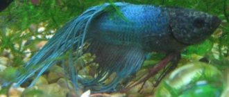
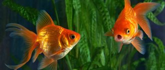
The family of cichlids or cichlids consists of more than 2 thousand species, distributed throughout the globe. Thanks to the variety of shapes and colors, and mainly to their mental abilities, these representatives of the perciformes order have earned the recognition of aquarium breed lovers.
Unfortunately, cichlids, like any aquarium inhabitants, are susceptible to various diseases. This is a test not only for patients, but also for owners. It’s terrible to see your pets suffer and waste away before your eyes. You have to urgently read the literature, consult with specialists, and look for the necessary medications.
The best treatment is prevention.
In order for your pets to remain healthy, first of all, you should take care of their habitat and proper care. But it also happens that, despite ideal living conditions and proper nutrition, they still manage to get sick. The main thing in this situation is to make a correct diagnosis and take timely measures.
Ichthyophthiriasis in cichlids: photos, symptoms, cause, treatment
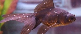
Ichthyophthiriasis is the most common disease of aquarium inhabitants. If cichlids rub against the ground and various objects, and small white bumps appear on the skin, reminiscent of semolina, you need to start treating the entire flock as soon as possible.
The causative agent of the disease is the Protozoa parasite, which matures in each tubercle - cyst, then leaves the body. The parasite lives autonomously from 7 to 10 days. All existing medicines destroy parasites not on the fish, but on those that have escaped into the open.
It is quite easy to treat ichthyophthyriasis in the early stages. The first thing you can do is raise the temperature to +30-32° C by adding about 2 g of salt per liter of liquid. This kills the parasites there. In this case, it is necessary to constantly change and saturate the water with oxygen.
The most common remedy is malachite green. Different forms are available, so you should read the instructions before use. To ensure the destruction of all parasites, the treatment course should not be less than 7-10 days. Malachite green does not cause side effects, so it can be used repeatedly.
The most effective drugs are Super Ick Cure, Faunomor and Costapur. They are harmless to vegetation and biofilters. They should be used in strict accordance with the instructions. Before starting the procedures, it is recommended to replace ¼ of the water in the container, and actively aerate during the process.
To prevent ichthyophthyriasis, it is recommended to quarantine new residents and clean live food purchased from sellers.
Diseases of aquarium fish caused by pathogens.
- Infectious - These are diseases that are caused by pathogens of plant origin: bacteria, viruses, fungi..
- Bacteria — Bacteria are predominantly single-celled organisms. The shapes are spherical, rod-shaped, convoluted, etc. Bacteria form spores, some are mobile, have flagella, feed, and, when the opportunity arises, attack fish. They attack fish in poor conditions. Bacterial diseases are common in aquariums.
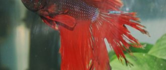
Fin rot (Aeromonas, Pseudomonas, Vibrio)
Photo: Fin rot (Aeromonas, Pseudomonas, Vibrio)In the initial stage, fish develop a barely noticeable bluish-white clouding of the edges of the fins and, in rare cases, the cornea of the eye. This is especially noticeable in small fish with transparent fins. Then the ends of the fin rays begin to fall off, and the edges of the fins become disheveled. In juveniles, the caudal fin disappears completely. And finally, ulcers form at the base of the destroyed fins, and the vertebral column is exposed, after which the fish dies.White-skinned (Pseudomonas dermoalba)
The disease begins to manifest itself through whitening of the skin around the dorsal fin and in the tail area. A characteristic sign of the disease: the fish stays near the surface of the water, often sticking its dorsal fin out of the water. The color fades or becomes yellowish (in red swordtails). If left untreated, the fish will die.
Fish tuberculosis, or Mycobacteriosis
Photo: Fish tuberculosis, or Mycobacteriosis. The symptoms of this disease are very diverse. Common symptoms for all types of fish: first, a sick fish loses its appetite, becomes lethargic, loses weight, its color turns pale, its fins are destroyed, its scales fall out; Sometimes there is darkening of the iris, bulging eyes, open ulcers, and black spots on the body. The listed signs can appear either singly or in combinations. Specific symptoms of the disease depend on the type of fish. In poeciliids, sick fish stay isolated from others and stop eating; exhaustion sets in, which is externally characterized by the fact that the back bends, the abdomen retracts, the eyes fall in, and bones protrude from under the skin and scales. In sick macropods, the skin is damaged and becomes red, the scales rise under the pressure of the liquid accumulated in the scale pockets and then fall out, sometimes an eyesore, bulging eyes occur, after which blindness occurs; the body becomes covered with black dots. In cockerels, the abdomen gradually increases, the skin begins to stretch and after 4-8 weeks becomes transparent; the fish become apathetic, but they move jerkily, often on their sides or belly up; stop eating. The zebrafish becomes bulging-eyed, and after some time the eyes fall out of their sockets, and the abdomen gradually increases in size.
Bulging fish eyes (Pop-eye)
Photo: Protruding eyes of fish (Pop-eye) The main symptom of this disease is the unnaturally bulging eyes of fish (telescopes and similar fish do not include this). Similar signs also occur with tuberculosis.
The fish looks bloated (Dropsy)
Photo: The fish looks bloated (Dropsy) The fish looks bloated and its scales stand on end like a Christmas tree.
Hole-in-the-head (Hexamitosis)
Photo: Hole-in-the-head (Hexamitosis)Small depressions form on the body, especially on the head. Often these ulcers are covered with a layer of mucus. Over time, these ulcers increase in size, leading to the death of the fish.
Tuberculosis
Photo: Tuberculosis The fish looks emaciated, is often affected by secondary bacterial and fungal infections, the eyes are bulging and ulcers form on the body and internal organs.
Ulcers on the fish's body
Photo: Ulcers on the fish’s body Ulcers appear on the fish’s body and internal organs.
Lepidorthosis
Photo: Lepidorthosis The diseased fish exhibits ruffled scales. In the initial stage, the scales rise only on a separate part of the body, then the ruffling spreads to the entire body, the scales fall out and the fish dies. Ruffled scales also occur when fish have tuberculosis and ichthyosporidosis. The rise of the scales occurs as a result of the formation under it of a small bubble filled with liquid - a pustule.Columnaria
Photo: Columnaria The fish develops a white mossy coating on the lips, and bruises with a filamentous border form on the body. When the gills are damaged, it rises to the surface, swallowing air.Bug eyes in fish
One or both eyes swell and protrude from their sockets. In extreme cases, the eye can literally “fall out” of its socket and become lost. Cloudiness of the entire outer surface of the eye is accompanied or preceded by exophthalmos. If exophthalmos is associated with a systemic infection (see below), then signs of this infection - for example, bloating - may be observed at the same time.
Corneal clouding in fish
One or both eyes are cloudy or have an opaque pupil.
Dropsy (ascites)
See description of the disease..
Malawi Bloat
See description of the disease..
Vibriosis
See description of the disease..
Flexibacter bacteria (synonyms - Chondrococcus, Myxobacteria)
See description of the disease..
- Fungal diseases — Fungi belong to a special kingdom of living organisms, combining both the characteristics of plants and animals, and have a higher organization than bacteria. Species pathogenic for fish include Vernal mycoses, Internal mycoses, and Branchiomycosis. Fungal diseases (mycoses) affect exclusively the surface of the body of fish in places of wounds, ulcers or due to bacterial infection. They are easy to notice by a coating of white mushroom threads, similar to cotton wool.
Dermatomycosis, or Saprolegnia
Photo: Dermatomycosis, or Saprolegnia On certain areas of the skin, fins, eyes, gills of fish in places of wounds and ulcers, white thin threads first appear, which soon form a cotton wool-like coating of white or light yellow color. The fins shorten, stick together and collapse, and may fall off. The fish becomes lethargic and inactive.Gill rot, or Branchiomycosis
Photo: Gill rot, or Branchiomycosis. Sick fish lose their appetite and have no reaction to external stimuli. Fish suffer from a lack of oxygen and therefore, rising to the surface, gasp for air. The gill covers are deformed. Pinpoint hemorrhages are observed on the gill filaments. The gills can have bright red, dark blue, pale and light areas. If severe damage occurs, the fish swims on its side and dies in this position.Ichthyophonosis, or Ichthyosporidiosis
Coordination of movements is impaired, the fish swims sluggishly, moves spasmodically, sometimes lies on the bottom, often on its side, and loses its appetite. Protruding eyes and their destruction in case of severe damage, trembling of the fins, raising of scales, knobby elevations, ulcers, dead areas on the body and fins, darkening of the color of the fish, and emaciation are also noted. Sometimes external signs are absent or barely noticeable.
Neon disease, or Plistiphora (Plistiphora hyphessobryconis)
Photo: Neon disease, or Plistiphora (Plistiphora hyphessobryconis) In neon, first the luminous stripe in certain parts of the body loses color, then becomes faded. In other fish, the color fades. Schooling fish stay alone. The fish stop taking food, swim restlessly all night, lose weight, hold their tail down at an angle of up to 60 degrees, make jerking movements to get out of this position, the spinal column is curved, the abdomen can become sunken, and sometimes the fins are destroyed.Mushrooms (fungus) on caviar
Photo: Mushrooms (fungus) on the eggs In the spawning aquarium and in the incubator, due to the large amount of organic matter and non-compliance with hydrochemical and temperature conditions, mold fungi of the genera Saprolegnia and Achlya settle on the eggs, the hyphae of which are visible in the form of thin white threads, which leads to the death of the eggs.
Cotton wool disease in fish
See description of the disease..
- Coelenterates — These include polyps, jellyfish, corals, ctenophores, which also have parasitic forms. The varieties are very difficult to distinguish and are found on different species of fish.
Oodinium
Photo: Oodinium At first the fish behave calmly, only occasionally scratching themselves on underwater objects. When examining the fish through a magnifying glass, on the body and fins, individual scales are bordered by small nodules in the form of a powdery rash of golden or gray color. Over time, the outer integument peels off in the form of flakes, and the interray tissue of the fins is destroyed. The fish swims in jerks. Mucus is secreted abundantly. The fish retain their appetite until death.Costia or Ichthyobodo
Photo: Costia or Ichthyobodo The affected fish first begins to rub against various objects, then refuses to feed. The introduction of the parasite causes severe irritation of the integument and the release of mucus. Dull bluish-grayish spots appear on the body of sick fish, which subsequently merge into a solid gray coating due to abundant mucus secretion. The most infected areas of the skin, where parasitic fungi settle, undergo decomposition. The gills turn pale and become covered with mucus, as a result of which breathing and gas exchange are disrupted, and the fish rise upward and swallow air. The fins are glued together. Pisces react poorly to external stimuli and become restless. In the absence of timely treatment, massive fish death may occur.Trichodinosis (Trichodina)
In the initial stage, the signs of the disease are weakly expressed: the fish stays near aeration bubbles, rubs against the ground, stones, and plants. When light is directed at the fish tangentially to the body, matte areas are visible. As the disease progresses, the body becomes covered with a whitish coating, which sometimes separates in the form of flakes. The gills also become covered with mucus and become pale in color. A sick fish refuses food, its breathing rate increases, it does not respond to external stimuli, makes oscillatory movements, and dies, turning on its side.
Chilodonella
Photo: Chilodonella Infected fish rub against rocks and plants and press their fins. Sometimes I lose my appetite. When viewed with the fish's head facing the observer, a bluish-matte coating is visible along the lateral line and slightly above it. In the worst case, the skin comes off in patches. When the gills are destroyed or damaged, there is no plaque, the fish is restless, often swims to the surface of the water, sometimes tries to jump out, then becomes lethargic. When gills become infected, massive fish deaths often occur.Glucose
Photo: Glugeosis A sick fish swims on its side, pineal-shaped protrusions are visible in various parts of the body, there are whitish and sometimes bloody opening spots and tumors, and one- or two-sided bulging eyes are observed. At autopsy, white nodules are visible on the affected organs (connective and muscle tissue, intestinal walls, kidneys, liver, gills, genitals, cornea of the eye). Microscopic examination of the nodules reveals accumulations of cysts.
- Flatworms (Plathelmintes) — There are parasitic and free-living forms of flatworms. Fish are usually parasitized by various forms of turbelaria, trematodes and cercomeromorphs.
Hematomas on the skin (Metacercariae)
Photo: Hematomas on the skin (Metacercariae) Due to the invasion of numerous cercariae, hematomas appear on the skin and muscles, and internal organs are exposed. During the invasion of cercariae, the fish swim restlessly, rub against rocks and breathe heavily. After the metacercariae in the fish calm down, the fish's behavior again becomes unnoticeable.Worms in the aquarium (Turbellaria, ciliated worms)
Photo: Worms in an aquarium (Turbellaria - Turbellaria, ciliated worms) The presence of ciliated worms in an aquarium can be easily determined: they are easily visible to the naked eye on glass, on stones and plants as white or light brown worms several millimeters in size.Parasites in the entrails of fish
Photo: Parasites in the insides of fish Signs are a swollen belly of the fish, slow swimming, sometimes worms are visible from the anus, sometimes they break through the body of the fish.
Eye flukes
See description of the disease..
"Black spots" in fish
One or more small dark spots of approximately circular shape, 1-2 mm in diameter, appear on the skin or fins of the fish. Even if these spots are present in large quantities, they do not have any harmful effect on the fish.
- Acanthocephalus - Of the 700 species of these worms, approximately 320 live in the intestines of vertebrates. Purely parasitic living acanthocephalans have a typical retractable proboscis equipped with a hook, with which they attach to the intestine of the victim. Acanthocephalans can enter the aquarium through food and with other fish.
- Threadworms (Nematoda) — .
Camallanus
The first sign of infection with these parasites is red-brown worms protruding from the anus of the fish. Severe infection can lead to spinal curvature and exhaustion.
Roundworms and threadworms (nematodes)
Mild infections may be asymptomatic. Severe infestations cause bloating and lethargy due to insufficient nutrient intake even though the fish are well fed. In extreme cases, spinal deformity may occur.
- Crustaceans (Crustacea) — .
Gyrodactylosis (Gyrodactylus)
Photo: Gyrodactylus (Gyrodactylus) At first, the affected fish stays near the surface of the water, sways, or makes oscillatory movements with the whole body, the fins are compressed. Then it begins to rub against underwater objects, individual parts of the body of a bluish-matte or gray color become visible, and the interray tissue of the fins is destroyed. The fish stops taking food, ulcers may appear on the skin, some slightly reddened areas, a bluish-white covering on the gills, and the eyes become cloudy. The gills are affected less frequently. In this case, their petals are destroyed. The fish suffocate, gather near the sprayer or on the surface of the water and swallow air. Without treatment, the fish gradually loses weight and dies.Dactylogyrus
A fish affected by the parasite behaves restlessly, stays near the surface, greedily swallowing air, does not take food, sways, rubs against objects. Its gills are covered with a thick layer of mucus, their color becomes faded due to anemia and takes on a mosaic-like character. The adjacent gill filaments grow together. In the acute form of the disease, the fish quickly die.
Lerneosis (Lernea)
Photo: Lerneosis (Lernea) The diseased fish sways, rubs against objects, ulcers with bright red edges are visible on the body, in which the crustacean can be seen with a magnifying glass.Argulus (Argulus foliaceus)
Photo: Argulus (Argulus foliaceus) This blood-sucking parasite, when it gets on a fish, causes an inflammatory process in the wound, which is characterized by copious mucus secretion on the body, redness and swelling. The fish sways and rubs against underwater objects.
Gill crustaceans
The behavioral signs are the same as those seen in gill diseases: shortness of breath, "coughing" and rapid breathing. When examining the gills, the female parasite (0.5-3.0mm in length) and her larval-like egg sacs (1-3mm in length) should be visible to the naked eye. Excessive mucus accumulates around the gills.
- Annelida (Annelida) — .
Fish leeches
See description of the disease..
Ichthyophthirvus multifiliis
Photo: Ichthyophthirvus multifiliis In diseased fish, small white dots first appear on the fins, and later on the entire body. The number of these points can increase rapidly after a few days. Before the dots appear, the fish behave restlessly, rubbing against the ground, plants, etc. When the gills are damaged, severe difficulty breathing occurs. In addition, other symptoms occur: loss of appetite, apathy, often approaching aeration bubbles.
Hexamitosis, or Octomitosis (Hexamita(Octomitus) trutta.)
The fish do not refuse food, but are very exhausted, swim spasmodically, the anus is inflamed, and sometimes the rectum and bladder partially fall out.
Ichthyophthirius, Ick, Ich, white spot
Photo: Ichthyophthirius (Ick, Ich, white spot)The most common disease. White cysts up to 1 mm in size appear on the fish. Sometimes they form clusters - as if the fish were sprinkled with sugar. Slaves scratch themselves on various objects. A secondary bacterial infection is often present.
Velvet desease (Oodinium)
Photo: Velvet desease (Oodinium)The body of the fish is covered with yellowish-gray dots. The dots resemble ichthyophthirius, but they are smaller in size and the fish looks as if sprinkled with gold dust. The fish scratches itself on various objects in the aquarium and breathes rapidly through its gills. Sometimes the skin comes off in strips.
Apiosoma (Apiosoma, Glossatella, Heteropolaria colisarum)
Photo: Apiosoma (Apiosoma, Glossatella, Heteropolaria colisarum) In small numbers, Apiosoma parasites are unlikely to cause any external signs, but clusters appear as whitish growths on the skin or fins, which can easily be confused with ichthyophthyriosis or lymphocystosis. Both diseases are widespread. The diagnosis can be confirmed by microscopic examination, but this is unlikely to be necessary. Ichthyophthyriasis can be distinguished by the rapid spread of infection. It quickly spreads throughout the fish’s body and spreads to other fish. Lymphocystosis causes the appearance of spherical neoplasms that form clusters like a bunch of grapes or cauliflower. Fluffy plaque or growths on places where the mucous membrane is damaged, which is difficult to confuse with a fungus, since the fungus has much longer threads.
Epistylis
Epistylis colonies look like small tufts of fungus. They usually appear on hard external surfaces such as gill covers and the tips of fin rays, often accompanied by accompanying signs of bacterial infection, or on infected wounds.
Guppy disease
Small white spots appear on the skin - colonies of protozoa. In guppies, these parasites sometimes accumulate around the eyes. The scales stick out to the sides like stubble (ruffles). Sometimes the muscles and internal organs are affected, and the disease can quickly lead to death. To make an accurate diagnosis, it is necessary to examine a scraping taken from the infected area of skin under a microscope.
Tetrahymena
See description..
Lymphocystis
Photo: Lymphocystis (Lymphocystis) Tiny white or gray nodules or flat growths, often black in color, on the skin and fins.
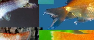
Fin rot in cichlids: photos, symptoms, cause, treatment
Quite often, a polluted environment causes fin rot. The disease is also provoked by stress and aggressive behavior of other inhabitants of the aquarium. The pathogen is transmitted from one individual to another through contact. Young cichlids are most susceptible to this. They most often die. Rot, as a rule, is a consequence of a systemic disease caused by harmful bacteria found in water, live food, and plants. New residents who have not completed quarantine can also bring them in.
Symptoms may overlap with characteristic signs of other diseases (bloating of the peritoneum, ulcers). Distinctive features of this particular disease may be:
- change in color of fins to dull blue along the edge with gradual expansion;
- the appearance of red spots, ulcers and stripes on them;
- decomposition of the caudal and pectoral fins, starting from the edge and moving to the base;
- Some individuals' eyes become cloudy.
If the disease is detected at an early stage, treating pets is not difficult. All that is needed is to replace a third of the liquid and heat it to 26° C. When there are inhabitants in the aquarium for whom this temperature is contraindicated, the procedure should be performed in a separate container. You need to dissolve 2-3 teaspoons of table salt in 5 liters of water and place the sick people there.
If after these procedures the condition does not improve, the use of medications is required.
The infection can be overcome by chloramphenicol (adult). You need to grind 1 tablet into powder and dissolve in 20 liters of liquid. Every third day you need to change 1/3 of the water in the container, and then dissolve a new dose of the medicine.
In a common tank, you can treat with streptocide (1.5 g of medicine per 10 l). First you need to dissolve it in a small container, then pour it into the aquarium.
Bicilin-5 is also good for getting rid of rot. Mix 250,000 units. funds with 10 liters of water in a small basin and put the patients there for 30 minutes. Duration of treatment – 6 days.
Biseptol-480 is used. The tablets are ground into powder and a solution is prepared before each session (1/8 tablet - per 5 l). Patients are treated for 7 days. The water is constantly saturated with oxygen.
To protect your pets from this disease, you need to change the water more often and be more attentive to the selection of aquarium neighbors.
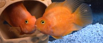
Description of the family
The cichlid family (lat. Cichlidae) belongs to the order Perciformes. There are about 200 species inhabiting the waters of Africa, Central and South America, and even in Southeast Asia there are 2 species. They mostly prefer fresh water, but some species can live in salted water.
Most members of the family have a tall body, slightly flattened laterally. The head is large relative to the body, the mouth has well-defined lips; in males, with age, a fatty growth appears on the forehead.
Mostly monogamous, they choose one partner and remain faithful to him for the rest of their lives. The spawning substrate can be varied: a flat pebble, a trench, a plant leaf. Some fish hatch eggs in their mouths. Males of these cichlids have rounded spots on their anal fin.
When the female pecks the spawned eggs for further incubation in her mouth, she tries to grab a spot on the anal fin of the gentleman, mistaking it for an egg, prompting the male to release milk into the oral cavity with the eggs.
Fertility is different for everyone:
- large cichlazomas can spawn about 2000 eggs;
- small fish that carry eggs in their mouths, no more than 100 eggs.
Cichlids are characterized by caring for their offspring; they protect and wash the eggs and larvae with their fins, and bite into food for the fry. As a rule, both parents care for the brood. The first eggs released are usually eaten, but subsequent eggs are hatched normally. If producers periodically eat caviar, then you can place the substrate with the eggs in a separate container, and allow aeration underneath.
They vary in whimsicality:
- Some cichlids, such as blackstripe and meeka, can be suitable even for beginners, you just need to provide them with enough space.
- But cichlids such as discus, apistogramma, and nannakars, on the contrary, require certain knowledge and experience from their owner.
They are not selective in feeding, and for the most part are predators, but there are species with a mixed diet or even herbivores.
All types of dry, live and frozen food can be used as food:
- tubifex;
- coretra;
- shellless shrimp;
- pieces of sea fish.
Herbivorous species and species with mixed nutrition can be fed:
- cucumber;
- food with the addition of spirulina algae;
- zucchini;
- carrot;
- spinach;
- scalded cabbage and lettuce leaves.
The behavior of cichlid fish is quite interesting, each individual has an individual character. Some cichlid breeders note that their pets are able to distinguish their owner from a stranger, “beg” for food, and sometimes even greet them home from work.
But, unfortunately, their interesting and complex nature gives rise to a lot of problems - most of them are aggressive territorial fish, in nature capable of attacking predators many times their size. Aggression especially increases during spawning, so it is advisable to provide new parents with a living space separate from their neighbors.
Bullying can be stopped by raising different types of cichlid fish together from juvenile age, providing sufficient aquarium volumes and a large number of necessary shelters and grottoes.
An exception:
- apistograms,
- discus,
- angelfish.
They have reduced aggression and love soft water. Small cichlids can be content with small aquarium volumes and do not dig up the soil, so they can be kept in a herbalist without fear for the beauty of the plants.
Hexamitosis in cichlids: photos, symptoms, cause, treatment
Hexamitosis (octomitosis, spironucleosis or hole disease) are several diseases with identical symptoms. They are caused by various intestinal parasites.
The first sign is loss of appetite. In the first stage, cichlids grab food but immediately spit it out. In the future, this leads to a complete refusal of food. Transparent thread-like discharge or discharge with food residues is the second symptom of the disease. Affected fish are darker than others and remain solitary. In patients, the stomach swells and the back dries out. Sometimes deep ulcers appear on the skin.
Depending on the circumstances, different methods and remedies are used:
- For bloating and loss of appetite, treat the aquarium environment with HexaEx once and give the fish medicinal food, which you can prepare yourself. To do this, you need to mix ½ tablet of NehaEx with 1 tbsp. a spoonful of semolina scalded and washed in a net;
- Bloating, complete lack of appetite - observe. If after 2 days the sick woman starts eating, try giving food with medicine, or a forced course of metronidazole or Sera bactopur direct. Continue feeding medicated food after appetite is restored;
- Clear stringy feces and lack of appetite. A course of metronidazole or Sera bactopur direct is required; if improvement occurs, medicinal food is given;
- Ulcers on the head and other signs – a complex of drugs (metronidazole, Sera bactopur direct) + medicated food.
If one individual is sick, you should not treat all of them at the same time. It is necessary to place the fish in another container, and feed the rest with medicinal food. Treat the aquarium with a single dose of HexaEx.
Treatment of pityriasis
When a patient is diagnosed with Gibert's disease, the dermatologist develops an individual treatment regimen to avoid dangerous complications. There is an opinion that pityriasis rosea will go away on its own in a few weeks. As a result, complications arise, and the patient comes to the dermatologist with an advanced form of the disease.
Drug treatment includes the following medications:
- antihistamines that relieve the patient from itching, swelling and redness on the body
- corticosteroid, desensitizing and antipruritic ointments. Medicinal compositions containing betamethasone, hydrocortisone, etc. are applied to the affected skin and lightly rubbed in. The medications eliminate rashes, get rid of peeling, and effectively restore the skin.
- drying agents containing zinc to accelerate skin healing
- Broad-spectrum antibiotics are indicated in cases where a bacterial infection is associated with pityriasis rosea. It is unacceptable to start taking antibacterial drugs on your own, since they are selected individually, taking into account laboratory tests.
- antifungal medications: drugs containing clotrimazole and other active substances are prescribed topically in the form of gels and ointments
- antiviral drugs containing acyclovir and other active components. Dermatological tests have confirmed that if antiviral drugs are prescribed in combination with antibiotics from the first days, the patient quickly recovers.
- neutral water-shaken preparations Dermatologists prescribe pharmacy talkers containing zinc oxide, menthol and anesthesin to patients with pityriasis rosea. These products relieve itching and pain in damaged areas and speed up recovery.
- iodine is an aggressive, but extremely effective remedy. Injured skin is treated with iodine in the morning and evening. Initially, the skin begins to peel off more actively, but then there are no extra scales left on it. Not all experts recommend that their patients cauterize damaged areas with iodine, since this drug can be harmful if used incorrectly.
Recommendations for patients
During the treatment period, the patient is recommended to adhere to the following recommendations:
- Follow a hypoallergenic diet: avoid nuts, citrus fruits, chocolates, honey, etc. Products containing artificial colors should be excluded from the menu. It is worth limiting the consumption of fried foods, carbonated drinks, fast food, strong alcohol and coffee.
- Limit water treatments within reasonable limits and give preference to the shower.
- Refuse to use aggressive hygiene and cosmetic products for the body. Gels and other detergents should not dry out the skin.
- Give preference to underwear made from natural fabrics.
- Moderate sunbathing - ultraviolet light helps the skin recover faster.
- Follow the recommendations of a dermatologist, apply to the skin only products recommended by a specialist.
- Folk remedies can be used only after consultation with your doctor.
Bug eyes (Exophthalmos) in cichlids: symptoms, cause, treatment
With poor care and deterioration of the chemical composition of the fluid, fish may develop bulging eyes or exophthalmos. Fluid accumulates inside or behind the eyeball, causing the eye to increase in size and protrude above the head. Typically, the disease affects one eye. It is considered non-contagious and disappears as suddenly as it appeared. Often exophthalmos is the result of an infection or fungal infection in a polluted environment.
Before you start treating underwater inhabitants, you need to check the quality of the aquatic environment. If this is the case, immediately begin cleaning it and change the water daily. In order to increase the immunity of fish, you should give food rich in vitamins and minerals. This will speed up the healing process.
In case of severe eye damage, magnesium sulfate is used. A sick cichlid is transferred to a container with a volume of up to 20 liters. 2 teaspoons of medicine are diluted in water. After a few days, the film disappears and the eye returns to normal.
Diseases of aquarium fish not caused by pathogens.
- Chemical origin — Diseases of chemical origin are all kinds of poisoning.
Fish poisoning with chlorine
Signs of severe poisoning appear and breathing becomes difficult. The gill filaments become covered with mucus and become light-colored, then the mucus appears on the skin. The fish rush around the aquarium, try to jump out of the water, then become lethargic and do not respond to external stimuli. Death comes suddenly.
Fish poisoning with ammonia, hydrogen sulfide, nitrates, nitrites
See description of the disease..
Metal poisoning of fish
Iron hydroxide poisoning damages the gills. As a result of heavy metal poisoning, the most important enzymes involved in metabolism are inhibited. As a result, a range of nonspecific symptoms may appear.
Fish poisoning by chemical industry products
See description of the disease..
Fish poisoning (general)
See description of the disease..
New Aquarium Syndrome
The same as for acute poisoning with ammonia and nitrites, in recently installed and not sufficiently matured aquariums.
- Physical origin — These are diseases associated with improper care: incorrect temperature, bad water, etc..
Lack of oxygen in fish (Anoxia)
The fish rise to the surface of the water and actively swallow air. Swallowing air, which continues for 1-2 hours, leads to protruding gills, then to “burning” of the fish’s gills with air and after a few days to the death of the fish. The decrease in oxygen content can also be judged by the behavior of snails, which rise from the bottom to the surface of the aquarium water. Keeping fish for a long time in conditions of lack of oxygen leads to poor appetite, slow growth, infertility, and weakened immunity.
Anemia (Anemia)
Sluggish movements, poor appetite, dystrophy are signs of anemia.
Acidosis (pH too low).
The disease does not appear immediately, but after some time. Fish become less active, swim less, but are more timid. At the same time, the appetite is preserved. The gill covers contract convulsively from time to time, and their movement slows down. The fish swims belly up or sideways. The color of the body becomes pale, sometimes milky white spots appear on it. Death occurs in thickets of plants, with their bodies curled up in a ring, mouth and gill covers closed. Even a slight change in acidity causes the fish to shudder convulsively, rush around the aquarium, and try to jump out of the water. This can lead to the death of fish. In some species of guppies, low pH causes the caudal fin to split.
The temperature is too high or too low
If the temperature is too high , the fish are extremely active, rushing up and down around the aquarium and trying to jump out of it. In addition, there is a lack of oxygen because In heating water, the oxygen content constantly decreases. If the temperature is low, there is lethargy in movements, loss of appetite, “swinging” of the whole body, slowing down or stopping the movement of the gill covers. Fish lie on the bottom, sometimes on their sides, or stand in one place, swaying their bodies. An even greater drop in temperature can lead to the death of fish. A drop in temperature can cause colds in the eggs or milt, in which the eggs and milt turn into a liquid mass, and the fish often die.
Gas embolism
Photo: Gas embolism External signs that help determine the presence of this disease in fish are small air bubbles on the inner surface of the aquarium glass, aquatic plants and decorations. These bubbles are a sure sign that the aquarium water is oversaturated with gases. And then the fish themselves can become covered with these bubbles, but this is not dangerous, it is dangerous if these bubbles accumulate under the skin and in the blood vessels. If this happens, it will lead to embolism and further death. The fish begin to swim on their sides and behave restlessly and fearfully. The fins and the whole body tremble convulsively. The movements of the gill covers slow down and then stop. Clouding of the cornea and ruffling of the scales may occur. The connective tissue of the fins may be destroyed.Inappropriate dH value
It is impossible to determine by external signs. There is only a general deterioration in the health of the fish and a weakening of the immune system.
Tumors in fish
Depends on the type of tumor. Melanosarcoma (tumor of pigment cells, malignant tumor) is easily recognized by its black color, which quickly spreads throughout the fish’s body. Other tumors are not accompanied by the appearance of pathogenic symptoms. If a tumor appears on the surface of the body, it can be easily recognized with the naked eye by its characteristic shape and often color. Sometimes the tumor is so large that it swells the fish's body. If the tumor is small and affects internal organs, it can only be detected at autopsy.
Alkaosis (pH too high)
The skin becomes dull and covered with mucus, the gills are damaged, mucus is released from them, the fins protrude, breathing becomes more frequent, loss of coordination of movements and convulsions occur. The fish are rushing around the aquarium, trying to jump out. As the disease drags on, clouding of the cornea of the eye occurs, leading to blindness. Eventually, death occurs, especially during the night hours.
Injuries in fish
Fresh injuries are quite easy to detect: they are visible to the naked eye. This is damage to the mucous membrane, scales, subcutaneous tissues and even muscles.
Asphyxia or suffocation of fish
When fish remain in water for a long time with insufficient oxygen content (less than 3 mg/l), oxygen starvation (anoxia) occurs, which ends in suffocation. The fish's breathing quickens, which is noticeable by the accelerated movement of the gill covers; they rise to the surface of the water and greedily gasp for air. The reduced oxygen content can also be judged by the behavior of sand snails, which emerge from the ground and can be seen on the glass of the aquarium and the leaves of plants.
Acidosis and alkalosis
Acute acidosis or alkalosis: excited behavior, rapid movement, jumping, which is often followed by death. Chronic acidosis or alkalosis: Less obvious signs include difficulty breathing and "coughing", excessive mucus production and itchy skin as a result of irritation caused by high acidity or alkalinity of the water. Problems with osmoregulation caused by chronic alkalosis can lead to bloating (see also section 6.3 - dropsy). Since the signs of acidosis, alkalosis and some other conditions are very similar, it may be necessary to determine the pH value to confirm the diagnosis. In cases of acidosis, if the pH value drops below 5, dark gray markings (iron deposits) may appear on the gills.
"Black chin" in fish
Small, irregularly shaped gray-black spots that first appear in the lower jaw area, and in severe cases spread along the underside of the fish's body from the head to the ventral fins, so that the entire lower part of the head and chest are mottled with gray-black spots. This is a skin disease that is apparently caused by environmental conditions. It affects cichlids, especially from lakes in the Rift Valley.
Problems associated with carbon dioxide (CO2)
Rapid breathing, loss of appetite. In the long term - slower growth in young fish. However, all these signs can be caused by other reasons.
Hypoxia (oxygen starvation) of fish
Accelerated breathing, hovering or swimming near the surface of the water, where the oxygen content is higher (in representatives of those species of fish for which such behavior is abnormal). In severe cases, breathing may become extremely difficult, the mouth is constantly open, and the gills become swollen. The fish usually lies on the bottom and is clearly suffering. Coordination of movements is impaired. Sometimes the intensity of the color increases, and the eyes become glassy and motionless. Note that some of these clinical signs are similar to those of acute poisoning and some infectious diseases that affect the gills and cause hypoxia.
Disease associated with the formation of gas bubbles
Lethargy, with no other signs of illness usually present. When examining the gills under a microscope, tiny bubbles can be found on the gill fibers. If gas bubbles form on glass and other surfaces in the aquarium, and the fish look unhealthy, then this disease should be suspected. In acute cases, bubbles may also appear on the fish themselves, sticking to the outer surface of the body. Death may occur as a result of the disease, and if exposure is not severe enough to cause death, it may cause brain damage. In spawning and nursery aquariums, affected eggs and fry may become buoyant, and the yolk sacs of the fry will visibly swell as they are filled with gas.
Hypothermia and overheating in fish
• Hypothermia: lethargy, lying on the bottom. When checking the water temperature, it is too low. If fish are exposed to refrigeration for too long, death may occur. • Overheating: rapid breathing, often shortness of breath, gills swollen and dilated. The fish hangs near the surface of the water, where the oxygen content is higher. Eventually exhaustion sets in, the fish lies on the bottom, followed by death from hypoxia. When checking the water temperature, it is too high.
Shock in fish
There are different ones, depending on the nature and severity of the lesion. Among them may be the following: a decrease in color intensity; rapid or slow breathing; lying on the bottom (a very common symptom) with periodic sudden movement to another place; in addition, fish hide among plants or other decorative objects. Sometimes shock manifests itself in the form of flight, and then the fish frantically rush around the entire aquarium up, down and along the walls, as if trying to find a way out. In severe cases, fish may lie on their sides or even belly up. Death from shock is quite common, especially among sensitive fish.
- The cause of the disease is in the food — These are diseases associated with improper feeding.
Obesity, high carbohydrate content, etc.
See description of the disease..
Gonadal cyst
Usually the disease is chronic. In this case, the abdomen with liquid or semi-liquid contents greatly increases in size due to the developed tumor. If treatment is not started on time, it will lead to infertility and death of the fish.
Constipation in fish
Lack of excrement is the main sign (other signs are nonspecific). It is often combined with loss of appetite. The fish may be lethargic and sometimes lie on the ground. In severe cases, the body swells and breathing becomes more rapid. Usually only one fish is affected.
Intestinal parasites in cichlids: symptoms, cause, treatment
The only disease that can seriously reduce a flock is cryptobiosis, caused by the parasite Cryptobia. Typical signs of infection in Malawian cichlids will be:
- darkening of the skin;
- bulging eyes;
- lack of appetite;
- lack of mobility;
- blindness.
In Tanganyika, only food refusal and a bloated abdomen are observed. The fry of lake cichlids die out almost completely within 1-2 weeks.
Regular ornidazole or secnidazole, sold in pharmacies, will help cope with intestinal infections. The medicine is thrown into a general aquarium once (1 tablet per 150 l). In advanced cases, repeat after 12 hours. Before the procedure, the fish should not be fed.
To give the disease a worthy rebuff, throw up to 5 tablets per 100 liters into a separate tank. The effect of the drug lasts 4 hours. During this time, the hungry fish will swallow the food along with the dissolved drug, and it will enter the intestines. If the cichlid's belly is swollen and it does not take food, the medicine is injected into the intestines with a syringe. In this case, the Canadian drug Tricaside is used. It is also added to frozen food (3 capsules per 1 kg) as a prevention of intestinal infections.
Dropsy in cichlids: photos, symptoms, cause, treatment
Excessive accumulation of fluid in the body, manifested by bloating of the body, is called dropsy or ascites. In addition, the excrement becomes like white strings, and the fish loses its appetite.
More often, this happens to cichlids living in poor conditions. Dropsy manifests itself as a sign of a bacterial disease (mycobacteriosis, nocardiosis) or a virus. The exact cause is difficult to determine. Therefore, it is better to isolate the sick individual and create an ideal environment for her. For bacterial etiology, antibiotics are used. But it is not always possible to cure.
Tuberculosis in cichlids: symptoms, cause, treatment
With this disease, caused by bacteria, small nodules appear in the internal organs of fish. When they break down, they form ulcers. In parallel, fin rot may occur. Tuberculosis may be suspected:
- general apathy;
- fading of color;
- decreased appetite;
- thinness;
- ulcers on the skin.
Fish become infected by eating the remains of deceased relatives. Tuberculosis in fish is incurable. Therefore, you should immediately remove all dead and suspicious fish, without waiting for the rest to start eating them.
Bloat (Malawi Disease) in Cichlids: Symptoms, Cause, Treatment
Malawi bloat is common among African cichlids of the Maingano type. The disease occurs in 2 stages:
Loss of interest in food. During this period, the disease is easy to cure. But it is quite difficult to notice unhealthy fish in a large school, and precious time is wasted.
The belly of the fish is greatly inflated, the body is covered with red spots and ulcers. White discharge, lethargy, and rapid breathing appear. Both individual signs and a combination appear. The last stage of the disease ends in death.
The exact causes of Malawi's bloating have not yet been established. With a certain degree of probability, we can say that protozoa that parasitize the intestines of fish are to blame.
Treatment with Metronidazole in a common space gives results only in the first stage. You need to mix 1 tablet with 200 liters of liquid. It should first be crushed and dissolved in warm water. The solution must be refilled daily for a week. Before each dose of the medicine, half the volume of the aquarium must be replaced.
Prevention of this disease is the best way to combat it. It is enough to maintain the optimal pH (water acidity) and dH (water hardness) values recommended for certain types of fish, and a balanced diet.
Neon disease in cichlids: photos, symptoms, cause, treatment
Most often, representatives of characins suffer from pathology, including ordinary neon, which is why neon disease got its name. In addition, cyprinids and cyprinids also get sick. It is caused by the protozoan parasite Pleistophora, which multiplies very quickly. Unsatisfactory living conditions contribute to this.
A characteristic sign of neon disease is a change in color. A white spot appears, quickly spreading to the middle of the body. The fish loses coordination and freezes in an inclined position with its tail up. After this comes death.
To date, no effective treatment methods have been invented. If the presence of pleistophora is confirmed, all aquarium inhabitants susceptible to the disease should be destroyed. Disinfect the tank, including all its contents.
Oodiniosis in cichlids: symptoms, cause, treatment
One of the most common pathologies caused by the protozoan parasite Oodinium pillularis is oodiniasis. It is equally dangerous for all types and ages of freshwater aquarium fish, including the Cichlid family.
The skin of sick fish becomes dull and becomes covered with a fine golden rash. Cell death occurs. The outer cover peels off. The fins stick together and break off, leaving broken rays. Mucus is released from the mouth, but appetite does not disappear. The fish moves in zigzags, trying to scratch itself on various objects.
Pathogens are introduced into the aquarium with new residents, vegetation and water from an infected container. They parasitize on the surface of the skin and fins. The most commonly affected species are cardinals, barbs and nanostomuses.
In order to cure fish, it is necessary to move them to quarantine at a temperature of about +25 ° C and treat them with Malachite greens with the addition of copper sulfate or Bicillin-5. To carry out disinfection in a common space, the brightness of the lighting is increased for two weeks, and the temperature is raised to 24-26° C. After this, the quarantine fish are returned to their original place of residence.
Causes
The causative agent of the disease hexamitosis is considered to be the flagellate parasite Hexamita salmonis (hexamita) - a teardrop-shaped microorganism with four flagella. The parasite affects the intestines and gall bladder of the fish. Hexamita reproduces by both division and the formation of cysts. Mature cysts leave the fish's body along with feces, infecting everyone in the aquarium.
It is quite possible that flagellates of other species similar to Hexamita salmonis parasitize aquarium fish. The fact is that hexamite was found on salmon fish, but did not cause damage to their skin. For aquarium fish, there have been no truly serious scientifically documented studies on this topic. And yet, quite effective methods have been developed to treat this disease.
It is traditionally believed that the cause of an outbreak of hexamitosis disease is poor care, and by correcting the maintenance, surrounding the fish with care and placing it in the most favorable conditions, this alone can provide effective treatment. This is wrong. Good living conditions will increase the fish’s body’s resistance, and the course of the disease can actually slow down and even stop. But in any stressful situation in this case, hexamitosis will resume. The fish must be seriously treated, applying therapeutic and preventive procedures to all other inhabitants of the home pond in order to be sure that the disease will disappear.
Return to content
Intestinal and gill worms in cichlids: symptoms, cause, treatment
Most often, in the intestines and gills of aquarium fish, in addition to microbes, representatives of the family of endoparasites - worms - settle. They cause loss of appetite, leading to general exhaustion. Gill worms cause fish to breathe more often, this is easily determined by the open gill covers. Patients float to the surface to grab as much air as possible with their mouths.
Each type of worm requires its own special measures. Medicines mixed into food have the most powerful effect on worms. The gill fluke is destroyed with organophosphorus preparations and formaldehyde. They also treat infection with crustacean parasites.
How to correctly diagnose the disease
The disease of fish is primarily manifested by staying in one place for a long time without moving, sinking to the bottom, or hiding under roots and decorative elements. There are many signs of disease in aquarium fish; as a rule, they have an external manifestation.
Sick fish
Signs of an unhealthy state of aquarium fish kept at home:
- Active breathing.
- The appearance of tufts and white spots on the fins.
- Changes in scale color.
- Red thin stripes on transparent fins.
- Changing the degree of pressing of the fins to the body.
- The appearance of a foreign body on the surface of the epidermis.
- The appearance of white dots.
- White round spots of different sizes.
- Abscess formation.
Early diagnosis of the occurrence of a disease in aquarium fish will make it possible to make the correct diagnosis as early as possible, choosing the right treatment and medications.
Carp eaters (carp lice) in cichlids: symptoms, cause, treatment
Karpovs can be seen with the naked eye. Adults (carp lice) are round in shape, about 4 mm in diameter. By sticking to the body, they drink the blood of the victim, leaving wounds into which various microbes then enter. Infected fish find no place for themselves, trying to rub against the soil, plants and design elements of the underwater home.
You can get rid of parasites using a 10% solution of ordinary potassium permanganate, placing the sick fish in it for 15-30 minutes. Having transplanted the inhabitants into a temporary home, the temperature in the common tank is maintained at +30° C for 3 weeks. After disinfection, the fish can be returned.
Carnivores end up in the aquarium along with food plankton, or with new residents who have not passed quarantine. Therefore, when planning to purchase pets, you should carefully examine not only them, but also their neighbors for bite marks. Interior decorations must be treated with an antiseptic, and it is better to refuse live food from natural bodies of water.
Compatibility
Typically, cichlid fish are aggressive only during the breeding season. At this time, they establish their territory, which is strictly prohibited for outsiders to enter. Naturally, the living area of the neighbors will be somewhat reduced, which can cause inconvenience for the entire population of the aquarium, so it is advisable to keep cichlids in spacious aquariums.
Note: It is not wise to keep several male parrot cichlids in one tank. The aquarium can be quite large and spacious, but only one male will take the lead and will chase the rest of the males and force them to hide in the thickets of plants.
Angelfish and parrots, as well as other calm and small cichlid fish, are well suited for living together: swordtails, mollies, platies, gourami, zebrafish, catfish, cockroaches, corydoras and other peaceful species with similar temperature preferences.
Lerneosis in cichlids: symptoms, cause, treatment
This dangerous disease is caused by worm-like copepods called lerna. Only females parasitize inside and on the surface of the body. They exhaust the victim by attaching themselves to the muscles and feeding on its blood. You can notice the crustacean by the 1 cm swellings protruding on the skin.
They get rid of the parasite using organophosphorus insecticides or ordinary tweezers. The wound left after the bite is disinfected.
Mycoses in cichlids: symptoms, cause, treatment
Parasitic fungi from the group of “water molds” create a fluffy coating on the body of the fish or on the eggs, filling everything and killing the victim. Infection is facilitated by unsatisfactory living conditions of underwater inhabitants and injuries. A fungal infection penetrates the wounds.
There are antifungal drugs available in pet stores that are effective in cases where mycosis has not spread too much. They also protect eggs from mold. Before use, you must carefully read the instructions.
Columnaria (Flexibacteriosis) in cichlids: symptoms, cause, treatment
An extremely contagious bacterial infection, Columnaria, is often a companion to fin rot. It usually affects the oral cavity, but can also appear on other parts of the body in the form of white spots that quickly become “moss”.
Flexibacteriosis develops in an unfavorable environment, stagnant water. Most often, migrants from other bodies of water who have experienced a sharp change in living conditions suffer.
Antibiotics are used to fight infection. They act quickly at the early stage of the disease. The deeper the infection penetrates into the body, the less effective the medicine is. For industrial breeding, phenoxyethanol is used.
Untreated hexamitosis will definitely appear again
You may be frightened by such a long and labor-intensive treatment, the number of procedures and potent drugs that may need to be used. But holey disease is not the case when you have to choose between the harm from the disease and the possible consequences of using medications. From this infection, fish die, die painfully, and at the same time infect others.
If the fish are not treated properly, the disease may freeze and “fall asleep” for a while. But even in its “dormant” form and under the most ideal conditions in the aquarium, it will cause great discomfort to your pets, and they will be contagious. Any negative factor: change of food, slight stagnation of water, temperature change - and a new outbreak will follow, in which it will be more difficult to save the fish.
Hexamitosis is too serious a disease; in its case, the potential harm from medications and long-term treatment still does not look as threatening as a new, larger epidemic and the death of your pets.
Return to content
Prevention of diseases in the aquarium
The best remedy for an epidemic in the aquarium world is preventive measures. Here are some simple tips that will help you avoid many troubles:
- Weekly replacement of 30% of the liquid volume with settled water;
- Cleaning the soil once a week;
- Cleaning rotten aquarium plants;
- Correct selection of diet and nutrition;
- Selection of quality feed and proper storage;
- Maintaining optimal temperature and lighting;
- Mandatory two-week quarantine for new residents;
- Transportation of pets in soft containers;
- Compliance with prescriptions and instructions when treating aquarium inhabitants;
- Do not install decorations in your home pond that emit toxic substances or objects that could injure fish;
- Aquarium accessories should not be used in other containers. Disinfect them every time after handling sick pets.
Prevention of dropsy
The best prevention of the disease is maintaining the necessary living conditions and minimizing stress. Find out at what temperature and pH of water the fish feel most comfortable in your aquarium. As a rule, these are the conditions of the natural habitat of this breed. A sharp change in temperature and water-salt conditions or their prolonged disturbance significantly increases the likelihood of disease in principle.
In addition, the quality of nutrition is of fundamental importance: the use of expired, dirty food, underfeeding or, most often, overfeeding is unacceptable. Water pollution also causes a decrease in immunity, so the incidence of dropsy is higher in places where the water in the aquarium is changed less frequently, that is, the excrement of the inhabitants accumulates in the soil.
Most non-aggressive fish require hiding places in the aquarium landscape, especially in the presence of more aggressive neighbors. The inability to hide is a big stress for a calm fish.
The principle of prevention is simple - the less stress a fish has to endure, the more successfully its body will cope with the threat of disease.