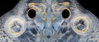
Fish are cold-blooded aquatic vertebrates that live in both salt and fresh water. Like mammals, fish have a closed circulatory system, meaning blood is always in the blood vessels unless they are damaged. Their circulatory system is quite simple. It consists of the heart and blood vessels. The heart is a primitive muscular structure that is located behind the gills.
The circulatory system of fish consists of the heart and blood vessels
Evolutionary development
According to research by biologists, it was in fish that for the first time among all living organisms the heart appeared as a full-fledged functioning organ. The skullless had only a pulsating vessel, the lancelets also lacked a heart, but the circulatory system itself became closed.
Today, such a development can be seen at the stage of ontogenesis, because fish embryos have only a vessel, which by the end of the incubation period is transformed into the heart muscle.
The circulatory system of the fish became closed, that is, from then on, the blood moved exclusively through the vessels, and the heart was divided into two chambers, equipped with a valve apparatus and a cardiac sac. This moment became the most important intermediate stage in the further development of the evolutionary chain.
After a while, animals began to go out onto land, and then they needed a more complex organ associated with pulmonary respiration.
Even in ancient times, lobe-finned fish acquired an additional respiratory organ, in which the main role is played by swim bladders, an incomplete cardiac septum and the second circle of blood circulation. Researchers have identified the rudiments of the third cardiac chamber in their structure. Some representatives of this unique group live on Earth today.
The neighbors of fish in their habitat - cephalopods - developed a gill heart millions of years ago. These are special extensions that create a pulsation along the path of the gill veins, through which venous blood flows. The number of such hearts always corresponds to the number of gills. Octopuses, cuttlefish and squids have two, while nautiluses have four.
What have we learned?
In a biology report for grade 7, it is important to tell what type of circulatory system is typical for fish. In short, fish were the first animals to develop a full two-chambered heart. It is quite weak, but ensures the continuous movement of blood through one closed circle of blood circulation, saturating organs and tissues with oxygen and getting rid of carbon dioxide and metabolic products.
Previous
BiologyVegetative propagation of plants by stem and leaf cuttings - types, methods and examples briefly (grade 6, biology)
Next
Biology Digestive system of reptiles - structural features of internal organs and functions briefly (grade 7, biology)
How many chambers do fish have in their heart?
The structure of the heart in fish includes two chambers. The first is a thin-walled atrium. It is in this section that the blood enters first. It is then pushed into the second chamber, which acts as a thick-walled ventricle. It is driven by the venous sinus (a sac with thin muscular walls) and the aortic cone (a pulsating muscular part with valves on the inner surface). Sometimes they are conventionally called the third and fourth chambers, which, of course, does not make the heart of fish similar to mammals and birds.
The four listed elements are located nonlinearly in the body of underwater animals: they form an S-shaped formation, where the aorta and ventricle are located below, and the vein and atrium are located above.
This is interesting: Fish have a very small heart: on average it occupies only 1%, while in mammals the volume reaches 4.7%, and in birds up to 16%.
Structure
Primitive fish have a two-chambered heart, which is conventionally divided into four segments:
- the first segment is a section called the venous sinus, which is responsible for receiving blood that gives oxygen to the body;
- the second segment is represented by the atrium with valves;
- the third segment is called the ventricle;
- the fourth segment is the aortic cone with several valves that pumps blood into the peritoneal aorta.
After the blood leaves the heart, it moves through the gills, where it is saturated with oxygen and flows into the spinal aorta, from where it is distributed to all tissues of the body.
In fish of the highest order, all segments are not located on the same line, but in the shape of the letter S, where the last two segments are located above the first two. This structure is characteristic of cartilaginous and lobe-finned fish. Bony representatives are distinguished by a weakly defined arterial cone, which is usually characterized as part of the aorta, and not the heart muscle.
What kind of blood is in the heart of a fish?
Venous and arterial blood flows in the body of representatives of ichthyofauna. Having given oxygen to the organs, venous blood flows through the liver to the heart, and from there to the gill filaments to again be saturated with vital gas. Oxygen-enriched blood is called arterial - it is the blood that goes into the dorsal aorta and from there it spreads through small vessels to other organs.
This is interesting: at the moment, fourteen blood groups have been identified in fish.
The color of arterial blood in fish is bright red, venous blood is dark cherry. The hue is given by red blood cells that have an oval shape (unlike the human disc-shaped one) and a nucleus. In addition to them, the blood contains:
- Plasma is a colorless liquid that carries blood cells;
- Hemoglobin is a protein that carries oxygen in red blood cells;
- Leukocytes are white cells that protect the body from pathogens and assist in the digestive system of fish;
- Platelets are cells that ensure the integrity of blood vessels by coagulating fluid;
In general, the chemical composition of the blood of fish is not very different from that of vertebrates: it includes organic and inorganic elements and metabolic products. But the percentage of the ratio of liquid tissue to the mass of fish among living beings is the smallest - from 2 to 7%.
A wide range of organs can create blood: gills, intestinal mucosa, kidneys, spleen, lymphoid organ located in the skull, and the heart of the fish. The role of an intermediary between blood and tissues in the body of fish is played by the lymphatic system - a set of vessels with a clear liquid (lymph).
Organ size
The size of the heart depends on the total body weight, so the larger the fish, the larger its “motor”. Our heart is compared to the size of a fist; fish do not have this opportunity. But as you know from biology lessons, small fish have a heart only a few centimeters in size. But in large representatives of the underwater world, the organ can reach even twenty to thirty centimeters. Such fish include catfish, pike, carp, sturgeon and others.
Mechanism of blood circulation
Compared to humans, fish have a very simple circulatory system, which consists of five main elements:
- Hearts with two chambers;
- Abdominal aorta;
- Dorsal aorta;
- Arteries and capillaries;
- Ven;
This is interesting: bony fish have no valves in their chambers, while the heart of cartilaginous fish (sharks and rays) has quite a lot of them. Thanks to them, animals do not experience strong pulse pressure, which can damage thin-walled gills.
Is there a swim bladder?
Cartilaginous fish do not have bladders, so swimming movements must be maintained constantly, even during sleep, otherwise they will sink to the bottom. The shark's caudal fin provides propulsion for swimming, the dorsal fin provides balance, and the pectoral fins are used to move up and down the depths.
The flattened body and posterior spine of stingrays make their swimming movements unique, unlike those of sharks. The large flattened body is fused with the pectoral fins, creating vertical waves when moving.
Chimeras use their pectoral fins when swimming, striking them simultaneously to move or alternately to change direction. This method is very effective, but is most common in bony fish.
Direction of blood circulation
Liquid tissue is transported through the circulatory system in only one direction - starting from the sinus venosus and ending with the conus arteriosus. One-way flow is ensured by valves that separate the chambers of the fish's heart.
In bony fish, which includes most species, venous blood that is not enriched, but cleared of toxins, flows through the aortic bulb into the abdominal aorta. Then it passes through four special channels - the afferent arteries - into the gills, where gas exchange occurs: oxygen penetrates into the blood, and carbon dioxide enters the environment.
From the efferent gill arteries, liquid tissue enters the epibranchial vessels: they form the head circle along the bottom of the skull and supply the brain of fish and other important organs of the head with substances. Next, the vessels form the dorsal aorta, located under the spine: smaller arteries and capillaries depart from it, which carry oxygen to the internal organs, muscles, and skin. From there, the blood returns through the capillaries to the veins.
The cardinal veins lead to the heart, where their ends join to form the ducts of Cuvier, which enter the sinus venosus, part of the heart muscle. The anterior vein carries current from the head organs of the nervous system of fish. The tail vein arises from the back of the body: it runs in the hemal canal directly below the artery.
In the area of the kidneys, this vein divides into a pair of portal veins. After filtration, the blood travels through the posterior cardinal veins to the heart, capturing red blood cells from the reproductive system along the way. All currents flow into the venous sinus, so that from there they can again begin pumping to the gills. Toxins are removed from the kidneys through the excretory system of fish.
In addition to the kidneys, the liver filters blood in the fish’s body. The portal veins of the liver take fluid tissue from the gastrointestinal tract, spleen and digestive glands, as well as a special organ - the swim bladder. Already purified blood exits through the capillaries into the paired hepatic veins and moves towards the heart.
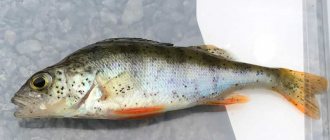
This is interesting: in some fish (for example, perch, carp and pike), the right renal portal vein is underdeveloped, so the organ cannot fully perform the cleaning function.
There are species that have significant deviations in the structure of the system. Thus, in cyclostomes, instead of a pair of four, seven afferent and efferent arteries, an unpaired epibranchial vessel, and the renal portal system and Cuvier’s ducts are completely absent. There is only one vein in the liver.
Cartilaginous animals have five afferent arteries in the gills, and efferent arteries - twice as many. In addition to those listed, they have subclavian vessels supplying the pectoral fins and brachial muscles, and lateral vessels in the abdominal cavity.
The already mentioned lungfishes have a special structure. Oxygenated blood is concentrated in the left side of the heart, and from there, through a pair of gill arteries, it enters the fish’s brain, head organs and dorsal aorta. The venous one, concentrated in the right half, leaves through a pair of posterior arteries and flows through the gills into the “lungs” - swim bladders.
When these animals breathe on the surface of the water, the blood is saturated in the air sacs and goes through the pulmonary veins to the left chambers. Additionally, abdominal and cutaneous veins were formed in the body of lungfish.
Important: capillaries are microscopic vessels that, thanks to their thin walls, transfer nutrients and oxygen to organ cells as quickly as possible. The intermediate part between capillaries and veins is called venules.
Anisimova I.M., Lavrovsky V.V. “Ichthyology” From-in Higher School. 1983
CHAPTER I STRUCTURE AND SOME PHYSIOLOGICAL FEATURES OF FISH
CIRCULOUS SYSTEM. FUNCTIONS AND PROPERTIES OF BLOOD
The main difference between the circulatory system of fish and other vertebrates is the presence of one circulatory system and a two-chambered heart filled with venous blood (with the exception of lungfishes and lobe-finned fish).
The heart consists of one ventricle and one atrium and is located in the pericardial sac, immediately behind the head, behind the last branchial arches, i.e., compared to other vertebrates, it is shifted forward. In front of the atrium there is a venous sinus, or venous sinus, with collapsing walls; Through this sinus, blood enters the atrium, and from it into the ventricle.
The expanded initial section of the abdominal aorta in lower fishes (sharks, rays, sturgeons, lungfishes) forms a contracting arterial cone, and in higher fishes it forms an aortic bulb, the walls of which cannot contract. Valves prevent blood from flowing back.
The blood circulation diagram in its most general form is presented as follows. Venous blood filling the heart, during contractions of the strong muscular ventricle, is directed forward through the bulbus arteriosus along the abdominal aorta and rises to the gills along the afferent branchial arteries. Bony fish have four on each side of the head, corresponding to the number of gill arches. In the gill filaments, blood passes through the capillaries and, oxidized and enriched with oxygen, is sent through the efferent vessels (there are also four pairs of them) to the roots of the dorsal aorta, which then merge into the dorsal aorta, which runs along the body back, under the spine. The connection of the aortic roots in front forms the head circle, characteristic of bony fish. The carotid arteries branch forward from the roots of the aorta.
From the dorsal aorta there are arteries to the internal organs and muscles. In the caudal region, the aorta becomes the caudal artery. In all organs and tissues, arteries break up into capillaries. The venous capillaries that collect venous blood flow into veins that carry blood to the heart. The tail vein, starting in the caudal region, enters the body cavity and divides into the portal veins of the kidneys. In the kidneys, the branches of the portal veins form the portal system, and after leaving them, they merge into paired posterior cardinal veins. As a result of the merger of the posterior cardinal veins with the anterior cardinal (jugular), collecting blood from the head, and the subclavian veins, bringing blood from the pectoral fins, two Cuvier ducts are formed, through which blood enters the venous sinus. Blood from the digestive tract (stomach, intestines) and spleen, passing through several veins, collects in the portal vein of the liver, the branches of which in the liver form the portal system. The hepatic vein, which collects blood from the liver, flows directly into the venous sinus (Fig. 21). In the dorsal aorta of the rainbow trout, an elastic ligament was found that acts as a pressure pump that automatically increases blood circulation during swimming, especially in the muscles of the body. The performance of this “extra heart” depends on the frequency of movements of the caudal fin.
Rice. 21. Scheme of the circulatory system of bony fish (according to Naumov, 1980): 1 – venous sinus, 2 – atrium, 3 – ventricle, 4 – aortic bulb, 5 – abdominal aorta, 6 – afferent gill arteries, 7 – efferent gill arteries, 8 – roots of the dorsal aorta, 9 – anterior jumper connecting the roots of the aorta, 10 – carotid artery, 11 – dorsal aorta, 12 – subclavian artery, 13 – intestinal artery, 14 – mesenteric artery, 15 – caudal artery, 16 – caudal vein, 17 – portal veins of the kidneys, 18 – posterior cardinal vein, 19 – anterior cardinal vein, 20 – subclavian vein, 21 – Cuvier’s duct, 22 – hepatic portal vein, 23 – liver, 24 – hepatic vein; vessels with venous blood are shown in black, vessels with arterial blood are shown in white
In lungfish, an incomplete atrial septum appears. This is accompanied by the emergence of a “pulmonary” circulation, passing through the swim bladder, transformed into a lung. The heart of fish is relatively very small and weak, much smaller and weaker than that of terrestrial vertebrates. Its weight usually does not exceed 0.33–2.5%, on average 1% of body weight, while in mammals it reaches 4.6%, and in birds even 10–16%.
Blood pressure (Pa) in fish is low - 2133.1 (skate), 11198.8 (pike), 15998.4 (salmon), while in the carotid artery of a horse - 20664.6.
The frequency of heart contractions is also low - 18–30 beats per minute, and it strongly depends on temperature: at low temperatures in fish wintering in pits, it decreases to 1–2; in fish that endure freezing into ice, the heart pulsates for this period stops.
The amount of blood in fish is relatively less than in all other vertebrates (1.1 - 7.3% of body weight, including 2.0-4.7% in carp, catfish - up to 5, pike - 2, chum salmon – 1.6, whereas in mammals – 6.8% on average).
This is due to the horizontal position of the body (there is no need to push blood upward) and less energy expenditure due to life in an aquatic environment. Water is a hypogravitational environment, i.e. the force of gravity has almost no effect here.
The morphological and biochemical characteristics of blood are different in different species due to the systematic position, characteristics of the habitat and lifestyle. Within one species, these indicators fluctuate depending on the season of the year, conditions of detention, age, sex, and condition of the individuals.
The number of erythrocytes in the blood of fish is less than that of higher vertebrates, and leukocytes, as a rule, are larger. This is due, on the one hand, to the reduced metabolism of fish, and on the other, to the need to strengthen the protective functions of the blood, since the environment is replete with pathogenic organisms. According to average data, in 1 mm3 of blood the number of red blood cells is (million): in primates – 9.27; ungulates – 11.36; cetaceans – 5.43; birds – 1.61–3.02; bony fish – 1.71 (freshwater), 2.26 (marine), 1.49 (anadromous).
The number of erythrocytes in fish varies widely, primarily depending on the mobility of the fish: in carp - 0.84–1.89 million / mm3 of blood, pike - 2.08, bonito - 4.12 million / mm3. The number of leukocytes in carp is 20–80, in ruff – 178 thousand/mm3. Fish blood cells are more diverse than those of any other group of vertebrates. Most fish species have both granular (neutrophils, eosinophils) and non-granular (lymphocytes, monocytes) forms of leukocytes in the blood.
Among leukocytes, lymphocytes predominate, accounting for 80–95%, monocytes account for 0.5–11%; among granular forms, neutrophils predominate – 13–31%; eosinophils are rare (in cyprinids, Amur herbivores, and some perches).
The ratio of different forms of leukocytes in the blood of carp depends on the age and growing conditions.
The total number of leukocytes in the blood of fish varies greatly throughout the year; in carp it increases in summer and decreases in winter during fasting due to a decrease in metabolic rate.
The blood is colored red by hemoglobin, but there are fish with colorless blood. Thus, representatives of the family Chaenichthyidae (from the suborder Nototheniidae), living in the Antarctic seas at low temperatures (<2°C), in water rich in oxygen, do not have red blood cells and hemoglobin in the blood. They breathe through the skin, which has a lot of capillaries (the length of capillaries per 1 mm2 of body surface reaches 45 mm). In addition, they have accelerated blood circulation in their gills.
The amount of hemoglobin in the body of fish is significantly less than that of terrestrial vertebrates: they have 0.5–4 g per 1 kg of body weight, while in mammals this figure increases to 5–25 g. In fast-moving fish, the supply of hemoglobin is higher than in sedentary (4 g/kg in anadromous sturgeon, 0.5 g/kg in burbot). The amount of hemoglobin in the blood of fish fluctuates depending on the season (in carp it increases in winter and decreases in summer), the hydrochemical regime of the reservoir (in water with an acidic pH value of 5.2, the amount of hemoglobin in the blood increases), nutritional conditions (carp raised on natural food and additional feed, have different hemoglobin levels). The acceleration of the growth rate of fish correlates with an increased supply of hemoglobin to their body.
The ability of blood hemoglobin to extract oxygen from water varies from fish to fish. Fast-swimming fish - mackerel, cod, trout - have a lot of hemoglobin in their blood, and they are very demanding of the oxygen content in the surrounding water. Many marine bottom fish, as well as eel, carp, crucian carp and some others, on the contrary, have little hemoglobin in the blood, but it can bind oxygen from the environment even with a small amount of oxygen.
For example, to saturate the blood with oxygen (at 16°C), pike perch requires a water content of 2.1–2.3 O2 mg/l; If there is 0.56–0.6 O2 mg/l in the water, the blood begins to release it, breathing becomes impossible and the fish dies.
For bream at the same temperature, the presence of 1.0–1.06 mg of oxygen in a liter of water is enough to completely saturate the blood hemoglobin with oxygen.
The sensitivity of fish to changes in water temperature is also associated with the properties of hemoglobin: as the water temperature rises, the body's need for oxygen increases, but the ability of hemoglobin to bind it decreases.
The ability of hemoglobin to bind oxygen and carbon dioxide is inhibited: in order for the oxygen saturation of the eel’s blood to reach 50% when the water contains 1% CO2, an oxygen pressure of 666.6 Pa is required, and in the absence of CO2, an oxygen pressure of almost half that is sufficient - 266. 6– 399.9 Pa.
Blood groups in fish were first determined on the Baikal omul and grayling in the 30s. It has now been established that group antigenic differentiation of erythrocytes is widespread; 14 blood group systems were identified, including more than 40 erythrocyte antigens. Using immunoserological methods, variability at different levels is studied; differences were identified between species and subspecies and even between intraspecific groups in salmon (when studying the kinship of trout), sturgeon (when comparing local stocks) and other fish.
Blood, being the internal environment of the body, contains in the plasma proteins, carbohydrates (glycogen, glucose, etc.) and other substances that play a large role in energy and plastic metabolism, in the creation of protective properties.
The level of these substances in the blood depends on the biological characteristics of fish and abiotic factors, and the mobility of the blood composition allows its indicators to be used to assess the physiological state.
Fish do not have bone marrow, which is the main organ for the formation of blood cells in higher vertebrates, or lymph glands (nodes).
Hematopoiesis in fish, compared to higher vertebrates, differs in a number of features: 1. The formation of blood cells occurs in many organs. The foci of hematopoiesis in fish are: gill apparatus (vascular endothelium and reticular syncytium, concentrated at the base of the gill filaments), intestines (mucosa), heart (epithelial layer and vascular endothelium), kidneys (reticular syncytium between the tubules), spleen, vascular blood, lymphoid organ (accumulations of hematopoietic tissue - reticular syncytium - under the roof of the skull). The prints of these organs show blood cells at different stages of development. 2. In bony fishes, hematopoiesis most actively occurs in the lymphoid organs, kidney and spleen, with the main hematopoietic organ being the kidneys (anterior part). In the kidneys and spleen, both the formation of red blood cells, white blood cells, platelets, and the breakdown of red blood cells occur. 3. The presence of both mature and young red blood cells in the peripheral blood of fish is normal and does not serve as a pathological indicator, unlike the blood of adult mammals. 4. Red blood cells, like other aquatic animals, have a nucleus, unlike mammals.
The spleen of fish is located in the anterior part of the body cavity, between the intestinal loops, but independently of it. This is a dense, compact dark red formation of various shapes (spherical, ribbon-like), but often elongated. The spleen quickly changes volume under the influence of external conditions and the condition of the fish. In carp, it increases in winter, when, due to reduced metabolism, blood flow slows down and it accumulates in the spleen, liver and kidneys, which serve as a blood depot; it is also observed in acute diseases. When there is a lack of oxygen, when transporting and sorting fish, or when fishing ponds, blood reserves from the spleen enter the bloodstream.
Changes in the size of the spleen in connection with periods of increased activity have been established in brook and rainbow trout and other fish.
One of the most important factors of the internal environment is the osmotic pressure of the blood, since the interaction of blood and body cells, water metabolism in the body, etc., largely depends on it.
The lymphatic system of fish does not have glands. It is represented by a number of paired and unpaired lymphatic trunks, into which lymph is collected from organs and along them is discharged to the terminal sections of the veins, in particular to the Cuvier ducts.
BackContentsNext
Heart rhythm
Contraction of the heart muscle occurs at a certain frequency, the speed of which depends on many factors:
- Biological characteristics of the species;
- Age;
- Health conditions;
- Ambient temperatures;
- Breathing movements;
Thus, in adult carp, the heart rate is on average 20-35 beats per minute, which is quite slow for fish. Especially in comparison with juvenile sturgeon, whose heart rate reaches 150 beats.
The most significant factor is temperature. As soon as the water gets colder, the fish’s heart begins to beat more and more slowly. During hibernation, a bream's heart beats to one beat per minute.
The main task of heart rate is to maintain a certain volume of blood flow corresponding to internal and external circumstances.
Electrical properties of the heart
Impulses in the heart muscle do not just happen. It is driven by cardiomyocytes - special organ cells that emit electrical impulses. In structure and functionality they are close to mammalian myocytes.
At rest, cardiomyocytes of teleosts and elasmobranchs are -70 mV, in hagfishes - about -50 mV. At the maximum potential level, the value changes from -50 mV to +15 mV. At the moment of depolarization of the membrane in the body, channels are excited through which sodium and calcium ions penetrate into the cells. At this moment, the heart enters the refractory stage - excitability drops, the state of the cells comes to a neutral level. On average, this phase in fish lasts about 0.15 seconds.
Subsequent membrane repolarization occurs due to the release of potassium ions from the cell. After which the potassium channels close, and the sodium channels do the opposite. So the cardiomyocyte potential returns to -50 mV.
In fish, myocytes are concentrated in specific parts of the heart muscle and together they form the conduction system. As in humans and other mammals, the initiation of systole in fish occurs at the synatrial node. But the function of a pacemaker (sinoatrial node) in fish is performed by all elements of the conduction system: the center of the ear canal and the node in the atrioventricular septum.
Important: the speed of achieving excitation in the cell in fish is lower than in higher animals, and it also varies within the organ of one individual.
What types are
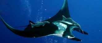
Cartilaginous fish, as you know, include sharks, rays, skates and chimaeras. There are about 600 different species of rays and 500 sharks on our planet. Stingrays are found in oceans all over the world. Most live on the bottom, and only a small part in open water. Among them are listed by name:
Manta ray (Manta birostris) feeds on plankton and small fish - the largest ray reaches 9 meters in width and weighs several tons.
Motoro - fresh water habitat, size up to 50 cm. It differs in color, there are many circles on the “cap”, which allows you to hide in the stones.
The common pipits (Dipturus batis) are the premier species of their genus, reaching up to 2.5 meters in length and having a lifespan of about 50 years. The smallest pipit is the star pipit (Raja stellata), which reaches a total length of 76 cm.
Chimeras are also known as ghost sharks and rats. Their closest living relatives are sharks, although evolutionarily they diverged from them nearly 400 million years ago. They lead an isolated lifestyle at great depths, at low temperatures. Like sharks and rays, chimeras have a cartilaginous skeleton, and the males have external reproductive organs (clasps) derived from the pelvic fins and used to introduce sperm into the female's body. They have a single external gill opening (there are different numbers of pairs), covered with a flap, like in bony fish, on each side of the body.
Male chimeras, unique among fish, possess an additional clamping organ, a tentacle on the forehead and in front of each pelvic fin. Many people mistakenly classify Protopterus (Protopterus) here because of the hidden gill sections. The place of this species in the class of bones.
A quarter of the world's fish, namely sharks and rays, will face extinction over the next few decades, according to the first study to systematically and globally assess the fate of cartilaginous orders. The study was conducted by the International Union for Conservation of Nature (IUCN for short) Shark Specialist Group (SSG), co-chaired by Nick Dulvey, Simon Fraser University's (SFU) Canada Research Chair in Marine Biodiversity. It shows that a quarter (249) of the 1,041 known species of sharks, rays and chimaeras worldwide fall into the critically endangered categories. The Mako shark is an agile and dangerous predator, based on which horror films are made. An adult can weigh 800 kg and measure up to 5 meters. In 2010, the representative was included in the Red List, the status is “vulnerable”. The knowledge gained will be useful not only for general development, but also for passing the OGE 2021.