Why is fungus dangerous?
Fungus is a contagious disease.
This fact alone brings a lot of problems, since its breeding environment (humid and warm) is found in many public places that people can visit daily (showers, swimming pools, gyms and beaches). Many people explain their ineffectiveness in treatment by the high cost and duration of treatment.
The fungus, without complete cure, during the severe stage can spread to internal organs. Even if all lesions are cured except one, it may lead to a return of the previous ones.
It has been scientifically proven that fungus reduces the quality of life, having a significant impact on social well-being, emotional well-being and health itself. According to the study, the quality of life in patients with fungal infection is reduced by half or even more, because in addition to natural physical inconveniences, itching, peeling, the patient has social problems, various fears, negative emotions that lead to mental problems.
The fungus can be transmitted hereditarily; more than a third of patients have relatives with a fungal infection. Therefore, there is a risk of transmitting the disease to children.
A fungal infection can cause other bacterial diseases, cause complications of existing skin and other diseases, for example, diseases of the endocrine system (diabetes mellitus), bronchial asthma, etc.
There are cases when neglected fungus led to deep mycosis. This is the germination of fungus on internal organs, complications and subsequent death of the patient. Therefore, if you have observed at least one of the symptoms, you should immediately contact a good specialist, and you should not self-medicate.
Bone diseases
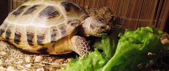
Signs of rickets, detachment and softening of the shell appear for the same reasons: due to insufficient absorption of calcium and other elements necessary for this process, as well as due to a deficiency of ultraviolet radiation. The main symptom of the pathology is thickening of the bones, while their structure and shape are disrupted.
It is difficult, but possible, to detect softening of bone tissue at the initial stage. If you carefully observe your pet, you will notice that the turtle has become lethargic and moves less than usual. This is exactly how reptiles with bone problems behave. If the shell has already softened, this is noticeable immediately: initially, the keratinized particles begin to peel off, and then the hard structure of the shield is replaced by a soft one, and perhaps even a change in shape.
You should consult your veterinarian before treating your turtle for bone disease. He will determine the causes of the disease and prescribe appropriate therapy. Most often it consists of taking vitamin and mineral preparations: Eleovita, Intravita, Multivita. If required, treatment is supplemented with other measures.
If you have bone diseases, your pet's diet needs to be adjusted. Redfish are given small fish with bones. In addition, for preventive purposes, it is recommended to place the reptile under a UV lamp, starting with 5-minute procedures, gradually increasing the time.
If the vet is out of reach...
Having noticed that the turtle has begun to molt, the owner should examine it and observe carefully. If, in addition to detachment, no other deviations from the norm are identified, the animal behaves actively, there is no reason for concern.
Having discovered that the shell is peeling off very much and even crumbling, you should resort to special treatment, which is prescribed based on the diagnosis. (The symptoms of various “shell troubles” were discussed above).
Fungus. Pharmacies sell antifungal drugs Nizoral or Lamisil. They should be used to lubricate the damaged areas of the shell at night for two weeks, having first very carefully cleaned off the exfoliated tissue.
The fungus can cause irreversible degeneration in the tissues of the turtle “armor”
Such tissues must be carefully cleaned and BetaisodonaR ointment applied to the ulcerated area.
In the case of a fungal disease, “therapeutic baths” are recommended for the amphibian - bathing in chamomile decoction, a weak solution of potassium permanganate or malachite greens. The latter, by the way, is used to disinfect aquarium water.
The disease in its unadvanced stage can be treated quite successfully. If the fungus reaches the internal organs, it can cause the death of the animal.
The shell of a land turtle can peel off as a result of dehydration. In this case, daily bathing of the animal is recommended.
As for rickets, which, as we found out, is the result of a deficiency of vitamin D and calcium, here we are most likely talking not so much about the treatment of the disease, but about its prevention.
So, in the warm season, it is recommended to take the aquaterrarium with the turtle out into the fresh air, while protecting the reptile from direct rays of the daylight.
In the autumn-winter period, turn on the UV lamp. In the autumn-winter period, the dwelling with the amphibian must be placed under a UV lamp three times a week - literally for five minutes.
Before the procedure, it is recommended to cover the turtle’s eyes with a plaster so as not to harm the lens and retina.
To replenish the level of calcium in the reptile’s body, shrimp rich in this element will not be superfluous in its menu. Since this pleasure is not cheap, you can treat your pet with fish and bones instead. Bone meal, crushed egg shells, and chalk are very useful.
If the disease is advanced, the veterinarian, as already mentioned, will prescribe vitamin injections.
Symptomatic treatment
Methylene blue will help eliminate white plaque (namely, get rid of plaque, but not the very reason that contributes to its appearance) - you can buy it at the pharmacy. Warm water (about 30⁰C) is poured into the aquarium and the drug is added to it until a light blue color appears. The turtle is allowed into the water for the whole day. In this case, it is imperative to prepare an area with a lamp under which the animal can warm up. At night the turtle is taken out of the water. Repeat for several days.
If desired, methylene blue can be replaced with malachite green, potassium permanganate or trypaflavin. You can also place the turtle in a bath of oak bark infusion for an hour.
Another way to quickly get rid of white spots is to wipe the shell with lemon juice.
But the above methods of therapy only relieve symptoms. To determine the causes of the pathology and cure the disease, you need to contact a veterinarian.
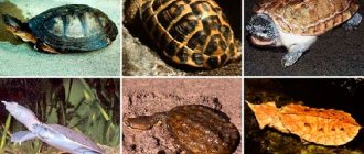
Bone diseases of red-eared turtles: symptoms and treatment (photo)
Rickets, detachment of parts of the shell and its softening are caused by the same reasons: poor absorption of calcium and other auxiliary microelements, as well as a lack of ultraviolet rays.
A symptom of rickets is a violation of the structure and shape of the bones, as a result of which thickenings appear on them. Determining the softness of bones at an early stage is quite difficult, but possible. To do this, you just need to take a closer look at your pet’s behavior. If your red-eared slider looks lethargic and doesn't move much, these are clear signs of a bone problem. As for the softening of the shell, its symptoms are immediately noticeable. First, its keratinized particles peel off, and then it becomes completely soft and even changes its shape.
Before treating bone diseases of red-eared turtles (see photo below), you should consult a veterinarian. He will help with advice and select the necessary complex of vitamins for the reptile, containing calcium and other useful microelements. These are, as a rule, “Eleovit”, “Intravit” and “Multivit”. If necessary, he may also prescribe additional treatment.
For these diseases, it will be useful to introduce small fish with bones into your pet’s diet. In addition, to prevent and treat any bone disease in red-eared turtles, it is helpful to place them under a lamp emitting ultraviolet rays from time to time.
Disease Prevention
This is a very important factor in the health of a domestic amphibian, and preventive measures are closely related to compliance with maintenance rules.
To prevent rickets and softening of the shell, the concentration of calcium in the body should be maintained. Especially for young cubs, bone meal should be added to their food daily. It will also be useful for adults (1 teaspoon once a week).
The risk of intestinal diseases is sharply reduced if you feed your pet only high-quality products. Greens and vegetables must undergo mandatory heat treatment. Don’t neglect special vitamin complexes for turtles.
The aquaterrarium should be kept clean, the water should be changed regularly, waste should be removed, and the required dose of UV radiation, temperature conditions, and normal ventilation should be provided.
Unfortunately, there are not so many veterinarians specializing in the treatment of amphibians. In some localities there are simply no veterinary clinics or offices. However, a recommendation from a veterinarian or an experienced turtle owner can always be obtained via the Internet. The main thing is to do this as quickly as possible and immediately begin proper treatment of your sick pet.
Various medications
Treatment tactics for heel fungus depend on the form of the disease. Usually a combination of tablets and external agents is selected. In the early stages, you can get by with only an ointment or solution. They help to effectively suppress microorganisms and lead to recovery. The course of treatment is two weeks. It must be completed to the end, even if the symptoms have disappeared.
Before applying the external product, the feet are steamed and washed with soap, and then wiped dry. The keratinized areas are carefully removed with a pumice stone or scraper. The medicine is applied to cleansed skin.
If necessary, the doctor prescribes tablets. Usually these are antibiotics. The most commonly used drugs are fluconazole, ketoconazole and others. The selection of medication must be done individually. They cannot be used during pregnancy and breastfeeding.
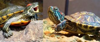
Fungus in the red-eared slider
The fungus on the shell of the red-eared slider is quite easily confused with a prolonged molting, in which the horny scutes are covered with white cobwebs. To clarify the diagnosis, determine the type of mycosis in the red-eared turtle and promptly prescribe treatment for your aquatic pet, you should contact a herpetologist or veterinarian.
The most common causes of fungal diseases in aquatic turtles are:
- diseases of bacterial, viral and parasitic nature;
- long-term uncontrolled treatment of animals with antibacterial drugs;
- frequent stress;
- cold water temperature in the aquarium, below 26C;
- lack of space for heating;
- mechanical damage to the shell;
- keeping the animal in salt water;
- unbalanced diet;
- hypo- and vitamin deficiency;
- lack of fluorescent and ultraviolet lighting;
- high water hardness;
- contact with infected relatives.
The combination of unfavorable factors against the background of decreased immunity, especially in the spring-autumn period, is an optimal environment for the proliferation of pathogenic fungi. Sometimes the cause of mycoses is the animal's prolonged stay on land, which results in drying out and the formation of cracks in the shell and skin.
Causes
The following can cause the appearance of white spots on the shell:
- hibernation, which weakens the body;
- prolonged exposure to cold water;
- inability to go onto land and bask under an ultraviolet lamp;
- long-term treatment with antibiotics;
- increased water hardness;
- poor nutrition, in which the body is deficient in vitamins and calcium;
- bad light;
- frequent stress;
- salty water;
- mechanical damage to the shell.
The shell also turns white in some diseases:
- Dermatomycosis - the causative agents of the disease are microscopic fungi Candida and Aspergillus, which always live on the body of reptiles, but manifest themselves only in cases where the immune system is weakened. At the same time, grayish-white cotton wool-like spots and granulomas appear in the folds of the skin and on the shell. After some time, the shell begins to deform and delaminate. In advanced cases, necrosis is observed.
- Saprolegniosis is caused by Saprolegnia fungi. The turtle's skin begins to turn red, peel and flake off, white “pimples” form on it, and later ulcers, which eventually begin to bleed. The shell becomes covered with a white coating, reminiscent of a cobweb, and begins to crumble. In reptiles, muscle tone weakens, activity decreases, and claws wear off. In advanced cases, paralysis of the limbs and sepsis are possible.
- Ulcerative-dissecting disease of the shell - first, the fungus Candida albicans enters the animal’s body through wounds and cracks formed on the shell, and later the bacteria Aeromonas hydropholy. The disease is accompanied by the formation of ulcers covered with whitish films, the number of which is constantly growing.
- Necrosis - pathogenic bacteria and fungi cause tissue death. As a result, light areas become noticeable on the limbs, head and shell, which darken after a while. If left untreated, the limbs swell, the nails and edges of the jaws begin to deteriorate and fall out. The animal dies from exhaustion or blood poisoning.
- Another reason for a white shell is a violation of molting, due to which the shell is covered with exfoliated skin that resembles a whitish film. In this case, there is no need for treatment. It is enough to remove skin flaps in a timely manner, normalize nutrition and living conditions.
Article on the topic: Eye diseases in chinchillas: suppuration, white discharge, cataracts and conjunctivitis
Types of deep mycosis
As noted above, there are about two dozen varieties of fungal infections that cause the development of deep mycosis. Some of them can be practically asymptomatic for a long time and become chronic. Others, on the contrary, have an acute course with pronounced symptoms.
Coccidioidosis is one of the most dangerous types of deep mycosis. Infection occurs through the respiratory route by inhaling dust containing fungal micelles. Once in the bloodstream, the causative agent of coccidioidosis spreads throughout the body, causing severe granulomatous inflammation of the skin, lungs, skeleton and brain. The clinical picture of coccidioidosis is similar to pneumonia, tuberculosis, meningitis, osteomyelitis, cellulitis and extensive abscesses.
Histoplasmosis is an extremely severe type of deep mycosis that is spread by airborne dust. Penetrating into the human body, it affects the mucous membranes, liver, spleen and bone marrow, and also provokes the formation of necrotizing erythematous spots on the skin.
Sporotrichosis is a fungal disease that often occurs in a chronic form. Infection occurs through deep damage to the skin, through the respiratory tract and from sick animals. At the beginning, the disease affects the skin, subcutaneous tissue and mucous membranes, and subsequently the infection spreads to the kidneys, liver and lungs.
Aspergillosis is a deep mycosis that affects the skin, sinuses, eyes, inner ear and lungs. It is transmitted through damage to the skin and the respiratory route, as well as from sick animals and birds. In severe cases, it causes the development of chronic ulcers and abscesses, bronchitis, pneumonia, otitis and panophthalmitis, up to complete loss of vision.
Cephalosporiosis is a dangerous disease that mainly affects the skin, subcutaneous tissue and musculoskeletal system. Infection occurs through injuries to the skin and mucous membranes. It is characterized by the development of ulcerative-erosive and gummous skin lesions and severe abscesses.
Cladosporiosis is a type of deep mycosis that most often affects the brain. The disease manifests itself in the development of multiple brain abscesses, which are accompanied by severe headaches and fever.
Mucorosis is a mold mycosis that can affect almost all internal organs and tissues, namely the skin, subcutaneous tissue, eyes, ears, bronchi, lungs, gastrointestinal tract, liver, kidneys and brain.
Mycoses of smooth skin
Among the widespread fungal diseases today, the most common are mycoses of smooth skin, such as microsporia, trichophytosis, lichen versicolor, mycosis of the feet (hands), and candidiasis. Sources of infection can be sick animals (cats, dogs, mouse-like rodents, cattle, etc.), as well as humans. In recent years, there has been an increase in the number of diseases caused by opportunistic fungi, among which superficial forms of candidiasis are most often recorded. Such a wide prevalence of these mycoses can be explained by the massive use of modern therapeutic agents, the environmental situation and other factors that reduce the body's defenses. One of the reasons for the significant prevalence of mycoses is the weakening of sanitary educational work in recent years. Due to insufficient awareness about the sources and ways of spreading infection, as well as adequate preventive measures, patients turn to the doctor late, and therefore mycoses become chronic, including in children suffering from mycoses of the scalp and smooth skin.
Microsporia is a fungal disease caused by various types of fungi of the genus Microsporum. In Russia, microsporia, which has spread over the last 50 years, is caused by a zoophilic fungus - fluffy microsporum (Microsporum canis), which parasitizes the skin of cats, dogs, and less often other animals. Infection from a sick person is observed in 2% of cases.
Epidemiology . Infection in 80-85% of cases occurs as a result of direct contact with a sick animal or through objects contaminated with the fur of these animals. Infection of children can also occur after playing in the sandbox, since the causative agent of microsporia is highly resistant to environmental factors and can remain viable in infected scales and hair for up to 7-10 years. Children often suffer from microsporia.
Clinic . After 5-7 days from the moment of infection, lesions appear on smooth skin, which can be observed on both open and closed parts of the body (children love to pick up animals and put them in bed with them). The lesions are round or oval in shape, pink or red, with clear boundaries, a raised ridge along the periphery, covered with blisters and thin crusts, with peeling in the center. The lesions are usually small, from 1 to 2 cm in diameter, single or multiple, sometimes merging. In 85-90% of patients, vellus hair is affected.
Treatment . If there are single foci of microsporia on smooth skin without damage to vellus hair, you can limit yourself to only external antifungal agents. The lesions should be lubricated with alcohol tincture of iodine (2-5%) in the morning, and sulfur-salicylic ointment (10% and 3%, respectively) should be rubbed in in the evening. You can rub the following antimycotics 2 times a day: mycozolon, mycoseptin, travogen or 1 time a day in the evening - mifungar cream, mycospor - until the clinical manifestations resolve. In case of multiple lesions of smooth skin and single lesions (up to 3) involving vellus hair, it is recommended to prescribe the antifungal antibiotic griseofulvin at the rate of 22 mg per 1 kg of the child’s body weight, in 3 doses after meals, in combination with exfoliating the stratum corneum of the epidermis in keratolytic lesions means (salicylic acid 3.0, lactic or benzoic acid 3.0, collodion up to 30.0). The lesions are lubricated with one of these products 2 times a day for 3–4 days, then 2% salicylic ointment is applied under compress paper for 24 hours, the detached scales of the stratum corneum of the epidermis are removed with tweezers and vellus hair is epilated. If during a control study carried out using a fluorescent lamp or microscope, affected hair is detected, the procedure is repeated. Detachment of the stratum corneum of the epidermis and manual hair removal of vellus hair can be carried out after using the “sealing” method. The lesions are sealed in a tile-like manner with strips of adhesive plaster for 2-3 days, this causes an aggravation of the process, which, in turn, facilitates hair removal.
The results of treatment for smooth skin microsporia are monitored using a fluorescent lamp or microscopic examination for fungi. The first control study is done after the resolution of clinical manifestations, then 3-4 days before the first negative test, and then after 3 days. The criteria for cure are resolution of the lesions, absence of luminescence and three negative tests on microscopic examination.
During the treatment, bed and underwear are disinfected: boiling in a soap-soda solution (1%) for 15 minutes (10 g of laundry soap and 10 g of caustic soda per 1 liter of water); ironing outerwear, furniture covers, and bedding five times with a hot iron through damp cloth.
Prevention. The main measure to prevent microsporia is compliance with sanitary and hygienic rules (you cannot use other people’s underwear, clothes, etc.; after playing with animals, you must wash your hands).
Trichophytosis is a fungal disease caused by various types of fungi of the genus Trichophyton. Trichophytons can be anthropophilic, parasitic on humans, and zoophilic, whose carriers are animals. Anthropophilic trichophytons include Trichophyton (Tr.) tonsuraus and Tr. violaceum, to zoophiles - Tr. mentagrophytes var gypseum and Tr. verrucosum.
Epidemiology. With superficial trichophytosis, caused by anthropophilic fungi, infection occurs through close contact with a sick person or indirectly through household items. Often children become infected from their mother, grandchildren from grandmothers suffering from a chronic form of the disease. The incubation period lasts up to a week. In zooanthroponotic trichophytosis, the sources of infection are sick animals: cattle, rodents. The highest incidence of this type of trichophytosis is recorded in the fall, which is associated with field work: it is at this time that the likelihood of infection through hay and straw increases. The incubation period ranges from 1–2 weeks to 2 months.
Clinic. On smooth skin with superficial trichophytosis, lesions can appear on any part of the skin - face, neck, chest, forearms. They have clear boundaries of a round or oval shape, with a raised ridge along the periphery of a bright red color; they are larger in size than in microsporia. The lesions are reddish-bluish in color, with peeling, nodules on the surface; in the chronic form, they develop on the skin of the buttocks, knee joints, forearms, less often the back of the hands and other parts of the body; the lesions do not have clear boundaries. Lamellar peeling is observed on the skin of the palms and soles. Vellus hairs are often affected.
With trichophytosis, caused by zoophilic fungi, the disease on the skin can occur in three forms: superficial, infiltrative and suppurative. Lesions are usually located on open areas of the skin. With a superficial form, they are round or oval in shape, with clear boundaries, a raised ridge along the periphery, on which bubbles, crusts, a pink center, and a bright red ridge are visible. The lesions are larger in size than with microsporia. Sometimes they are located around natural openings - eyes, mouth, nose. In the infiltrative form, the lesions rise above the skin level and are accompanied by inflammatory phenomena - infiltration. The suppurative form is characterized by the development of tumor-like formations, bright red in color, covered with purulent crusts due to the addition of a bacterial infection. When the lesion is compressed, pus is released from the hair follicles and pain is noted. The disease is accompanied by a violation of the general condition, sometimes the temperature rises. After the resolution of clinical manifestations, cicatricial atrophy of the skin remains at the site of former lesions. Clinical forms of zooanthroponotic trichophytosis can transform into one another.
Diagnostics. The diagnosis of trichophytosis is established on the basis of the clinic and when the fungus is detected during microscopy of pathological material, and the type of pathogen is determined using cultural examination.
Treatment. Treatment is carried out with antimycotics for external use. The lesions are lubricated with tincture of iodine (2-5%) during the day, and sulfur-salicylic ointment (10% and 3%, respectively) or mycoseptin is rubbed in in the evening. You can carry out monotherapy with ointment or cream (kanison, mifungar, mycozoral, mycospor (bifosin), exoderil, mycozoral, etc. In the infiltrative form, 10% sulfur-tar ointment is prescribed to resolve infiltration 2 times a day. Treatment of the suppurative form of trichophytosis begins with removing crusts from the affected area using bandages with 2% salicylic ointment, which are applied for several hours. After removing the crusts, vellus hair is epilated. Then apply lotions with solutions that have a disinfectant and anti-inflammatory effect (furacilin 1:5000, rivanol 1:1000 , potassium permanganate 1:6000, ichthyol solution (10%), etc.). As a result of this treatment, the hair follicles are freed from pus, inflammatory phenomena are reduced. Next, sulfur-tar ointment (5-10%) is prescribed for resorption of the infiltrate (5-10%) in the form of rubbing or under wax paper.After the infiltrate has resolved, antimycotics are used for external use (see superficial form of trichophytosis).In cases where vellus hair is affected in lesions on smooth skin, the stratum corneum of the epidermis is detached, followed by hair removal. To do this, you can use salicylic collodion (10-15%), milky-salicylic-resorcinol collodion (15%). If there is no effect, griseofulvin is prescribed orally at a daily dose of 18 mg per 1 kg of body weight, in 3 divided doses after meals daily until a negative test for fungi, then every other day. As an alternative method, terbinafine (Lamisil, Exifin) can be prescribed to adults 250 mg (1 tablet) once a day after meals every day, children weighing up to 20 kg - 62.5 mg, from 20 to 40 kg - 125 mg , over 40 kg - 250 mg in combination with antimycotics for external use.
The criteria for cure for trichophytosis are resolution of clinical manifestations and three negative fungal test results at three-day intervals.
Prevention. Prevention of trichophytosis depends on the type of pathogen. With superficial trichophytosis caused by anthropophilic fungi, the main preventive measure is to identify the source of infection, and it can be children suffering from superficial trichophytosis, or adults suffering from a chronic form of the lesion. In recent years, cases of chronic trichophytosis have been observed in middle-aged and older children. For suppurative trichophytosis, preventive measures are carried out jointly by medical workers, epidemiologists and veterinary services.
Mycosis of the smooth skin of the feet (hands). In a number of countries, up to 50% of the population suffers from mycosis of the feet. This disease is more common in adults, but in recent years it has often been observed in children, even infants.
Etiology. The main causative agents of mycosis of the feet are the fungus Trichophyton rubrum (T. rubrum), which is isolated in almost 90% of cases, and T. mentagrophytes var. interdigitale (T. interdigitale). Damage to the interdigital folds, which may be caused by yeast-like fungi, is recorded in 2-5% of cases. The anthropophilic fungus Epidermophyton floccosum is rarely isolated in our country.
Epidemiology. Infection with mycosis of the feet can occur in the family through close contact with a patient or through household items, as well as in a bathhouse, sauna, gym, or when using someone else's shoes and clothes.
Pathogenesis. The penetration of fungi into the skin is facilitated by cracks and abrasions in the interdigital folds caused by sweating or dry skin, abrasion, poor drying after water procedures, narrowness of the interdigital folds, flat feet, etc.
Clinic. Clinical manifestations on the skin depend on the type of pathogen and the general condition of the patient. The T.rubrum fungus can cause damage to the skin of all interdigital folds, soles, palms, dorsum of the feet and hands, legs, thighs, inguinal-femoral, intergluteal folds, under the mammary glands and axillary area, trunk, face, and rarely the scalp. The process may involve vellus and long hair, nail plates of the feet and hands. When the skin of the feet is affected, there are 3 clinical forms: squamous, intertriginous, squamous-hyperkeratotic.
The squamous form is characterized by the presence of peeling on the skin of the interdigital folds, soles, and palms. It can be flour-shaped, ring-shaped, lamellar. In the area of the arches of the feet and palms, an increase in the skin pattern is observed.
The intertriginous form is the most common and is characterized by slight redness and peeling on the lateral contact surfaces of the fingers or maceration, the presence of erosions, superficial or deep cracks in all folds of the feet. This form can transform into dyshidrotic, in which vesicles or blisters form in the area of the arches, along the outer and inner edges of the feet and in the interdigital folds. Superficial blisters open with the formation of erosions, which can merge, resulting in the formation of lesions with clear boundaries and oozing. When a bacterial infection is attached, pustules, lymphadenitis and lymphangitis occur. In the dyshidrotic form of mycosis, secondary allergic rashes are observed on the lateral and palmar surfaces of the fingers, palms, forearms, and shins. Sometimes the disease becomes chronic with an exacerbation in spring and summer.
The squamous-hyperkeratotic form is characterized by the development of foci of hyperkeratosis against the background of peeling. The skin of the soles (palms) becomes reddish-bluish in color, and pityriasis-like peeling is noted in the skin grooves, which extends to the plantar and palmar surfaces of the fingers. Pronounced ring-shaped and lamellar peeling may be detected on the palms and soles. In some patients, it is insignificant due to frequent hand washing.
In children, lesions of smooth skin on the feet are characterized by fine-plate peeling on the inner surface of the terminal phalanges of the toes, usually 3 and 4, or there are superficial, less often deep cracks in the interdigital folds or under the toes, hyperemia and maceration. On the soles, the skin may not be changed or the skin pattern may be enhanced; sometimes ring-shaped peeling is observed. Subjectively, patients are bothered by itching. In children, more often than in adults, exudative forms of lesions occur with the formation of blisters and weeping eczema-like lesions. They appear not only on the feet, but also on the hands.
Rubrophytia of smooth skin of large folds and other areas of the skin is characterized by the development of lesions with clear boundaries, irregular outlines, with an intermittent ridge along the periphery, consisting of merging pink nodules, scales and crusts, with a bluish tint (the color is bluish-pink in the center) . On the extensor surface of the forearms and shins, rashes can be located in the form of open rings. Lesions with nodular and nodular elements are often observed. The disease sometimes occurs as an infiltrative-suppurative trichophytosis (more often in men when localized in the chin area and above the upper lip). Foci of rubrophytosis on smooth skin can resemble psoriasis, lupus erythematosus, eczema and other dermatoses.
The fungus T. interdigitale affects the skin of the 3rd and 4th interdigital folds, the upper third of the sole, the lateral surfaces of the foot and toes, and the arch of the foot. This mushroom has pronounced allergenic properties. With mycosis of the feet caused by T. interdigitale, the same clinical forms of damage are observed as with rubrophytosis, but the disease is often accompanied by more pronounced inflammatory phenomena. In the dyshidrotic, less often intertriginous form, large blisters may appear on the skin of the soles and fingers, along with small blisters; in the case of bacterial flora, with purulent contents. The foot becomes edematous, swollen, and pain appears when walking. The disease is accompanied by an increase in temperature, deterioration of health, development of allergic rashes on the skin of the upper and lower extremities, torso, face, enlargement of the inguinal lymph nodes; the clinical picture is similar to that observed with eczema.
Diagnosis. The diagnosis is established on the basis of clinical manifestations, detection of the fungus by microscopic examination of skin flakes and identification of the type of pathogen by cultural examination.
Treatment. Treatment of mycosis of the smooth skin of the feet and other localizations is carried out with antifungal agents for external use. For squamous and intertriginous forms of lesions on the feet and other areas of the skin, medications are used in the form of a cream, ointment, solution, spray; you can combine a cream or ointment with a solution, alternating their use. Currently, the following medications are used to treat this disease: exifin cream, mycozoral cream, nizoral cream, canizon cream and solution, mycozon cream, mycospor (bifosin) cream, mifungar cream, lamisil cream and spray, mycoterbin cream. These drugs are applied to cleansed and dried skin once a day, the average duration of treatment is no more than 2 weeks. Antimycotics such as travogen, ekalin, batrafen, mycoseptin, mycozolon are used 2 times a day until clinical manifestations resolve, then treatment is continued for another 1-2 weeks, but once a day - to prevent relapse. In nodular and nodular forms of rubrophytosis, after relieving acute inflammatory phenomena using one of these ointments, sulfur-tar ointment (5-10%) is prescribed in order to further resolve the clinical manifestations. For intertriginous and dyshidrotic forms (the presence of only small blisters) of mycosis of the feet, drugs with a combined effect are used, which, along with an antifungal agent, include a corticosteroid, for example mycozolon, travocort, or a corticosteroid and an antibacterial drug - triderm, pimafucort.
In case of acute inflammatory phenomena (wetting, the presence of blisters) and severe itching, treatment is carried out as for eczema: desensitizing agents (intravenous or intramuscular administration of calcium chloride solution (10%), sodium thiosulfate solution (30%), calcium gluconate solution (10%) or calcium pantothenate orally; antihistamines. For external medications, at the first stage of therapy, lotions are used (2% boric acid solution, potassium permanganate solution 1:6000, 0.5% resorcinol solution), 1-2% aqueous solutions of methylene blue or brilliant green, fucorcin. Then they switch to pastes - boron-naphthalan, ichthyol-naphthalan, ACD paste - F3 with naphthalan, if complicated by bacterial flora - lincomycin (2%). At the 2nd stage of treatment after resolution of acute inflammatory phenomena, the above-mentioned antimycotic agents are used.
A drug such as Triderm, which contains, in addition to an antimycotic (clotrimazole 1%), a broad-spectrum antibiotic (gentamicin sulfate 0.1%) and a corticosteroid (betamethasone dipropionate 0), can quickly and effectively eliminate the symptoms of inflammation and itching in the presence of both fungal and bacterial infections. .05%). The presence of Triderm in 2 dosage forms - ointment and cream - makes it possible to use it for different types and at different stages of the pathological process.
If external therapy is ineffective, systemic antimycotics are prescribed: itraconazole in a continuous regimen of 200 mg per day for 7 days, then 100 mg for 1-2 weeks; terbinafine (Lamisil, Exifin) 250 mg once a day every day for 3-4 weeks; fluconazole (150 mg once a week for at least 4 weeks).
Prevention. To prevent foot mycosis, it is necessary to observe, first of all, the rules of personal hygiene in the family, as well as when visiting a bathhouse, sauna, swimming pool, gym, etc.; disinfect shoes (gloves) and linen during the treatment period. After visiting a bathhouse, swimming pool, sauna, to prevent mycosis of the feet, apply Daktarin spray powder to the skin of the interdigital folds and soles.
Tinea versicolor is a fungal disease caused by Malassezia furfur (Pityrosporum orbiculare), a yeast fungus. Lichen versicolor is quite widespread in all countries; young and middle-aged people suffer from it.
Etiology. Malassezia furfur as a saprophyte is found on human skin and, under favorable conditions, causes clinical manifestations.
Pathogenesis. Factors contributing to the development of the disease have not yet been precisely established, however, lichen versicolor is more common in people suffering from excessive sweating, changes in the chemical composition of sweat, diseases of the gastrointestinal tract, endocrine pathology, vegetative-vascular disorders, as well as immune deficiency .
Clinic. The disease is characterized by the presence of small spots on the skin of the chest, neck, back, abdomen, less often the upper and lower extremities, axillary and inguinal-femoral areas, on the head; the spots are initially pink in color and then become light and dark brown; Slight peeling is also observed, sometimes it can be hidden and can only be revealed by scraping. The rashes often merge, forming large areas of damage. After tanning, as a rule, white spots remain as a result of increased flaking. The disease is characterized by a long course with frequent exacerbations.
Diagnosis. The diagnosis is made on the basis of clinical manifestations, when the pathogen is detected in skin flakes during a microscopic examination and in the presence of a characteristic yellow or brown glow under a Wood's fluorescent lamp, as well as a positive iodine test.
Treatment. Currently, there is a sufficient selection of antifungal drugs for topical use that have a pronounced antifungal effect against the causative agent of lichen versicolor. These include imidazole and triazole derivatives, allylamine compounds. During the treatment of the disease, the following is used: exifin cream (applied to cleansed and dried skin in the affected areas 2 times a day for 7-14 days, if necessary, after a 2-week break, the course of treatment can be repeated), nizoral cream, mycozoral ointment, cream and canizon solution, mycozon cream, mifungar cream (prescribed once a day, duration of treatment is 2-3 weeks); lamisil cream and spray; nizoral shampoo (for three days, apply to the affected areas of the skin for 3-5 minutes and wash off in the shower). For common, often recurrent forms of lichen versicolor, systemic antimycotics are more effective: itraconazole (prescribed 100 mg once a day for two weeks, then take a two-week break, repeat the course of treatment if necessary), fluconazole (150 mg once a week within 4-8 weeks). During treatment, it is necessary to disinfect the patient’s clothes, hats, underwear and bed linen by boiling in a 2% soap-soda solution and ironing with a hot iron while wet. Family members of the patient should also be examined.
Prevention. To prevent recurrence of mycosis, it is necessary to use nizoral shampoo. Treatment should be carried out from March to May once a month for 3 days in a row.
Smooth skin candidiasis is a fungal disease caused by yeast-like fungi of the genus Candida.
Etiology. The pathogens are opportunistic fungi that are widely distributed in the environment. They can also be found on the skin and mucous membrane of the mouth, digestive tract, and genitals of a healthy person.
Epidemiology. Infection from the external environment can occur with constant fractional or massive infection with fungi.
Pathogenesis. Both endogenous and exogenous factors can contribute to the occurrence of candidiasis. Endogenous factors include endocrine disorders (usually diabetes mellitus), immune deficiency, severe somatic diseases and a number of others. The development of the disease is possible after the use of a number of modern medications: broad-spectrum antibiotics, immunosuppressive and hormonal drugs. The occurrence of candidiasis in the interdigital folds of the hands is facilitated by frequent contact with water, as this develops maceration of the skin, which is a favorable environment for the introduction of the pathogen from the external environment.
Clinic. On smooth skin, small folds on the hands and feet are more often affected, less often - large ones (inguinal-femoral, axillary, under the mammary glands, intergluteal). Lesions outside the folds are located mainly in patients suffering from diabetes mellitus, severe general diseases, and in infants.
In some patients, the disease begins in small folds of the skin with the formation of small, barely noticeable blisters on the lateral contacting surfaces of hyperemic skin, the process gradually spreads to the area of the fold, then peeling, maceration appears, or immediately shiny eroded surfaces of a deep red color with clear boundaries appear, with peeling of the stratum corneum of the epidermis along the periphery. The 3rd and 4th interdigital folds on one or both hands are most often affected. The disease is accompanied by itching, burning, and sometimes pain. The course is chronic, with frequent relapses.
In large folds, the lesions are dark red in color, shiny, with a moist surface, with a strip of exfoliating stratum corneum of the epidermis, occupying a significant surface, having clear boundaries and irregular outlines. New small erosions appear around large foci. In children, the process of large folds can spread to the skin of the thighs, buttocks, abdomen, and torso. Painful cracks sometimes form deep in the folds.
Candidiasis of smooth skin outside the folds has a similar clinical picture.
Diagnosis. The diagnosis is made on the basis of a typical clinic when a fungus is detected in scrapings from skin flakes during a microscopic examination.
Treatment. Limited and sometimes widespread acute forms of smooth skin lesions, especially those that developed during therapy with antibacterial drugs, as a rule, are easily treated with local antimycotic agents in the form of a solution, cream, ointment and can resolve even without treatment after discontinuation of antibiotics.
For candidiasis of smooth skin of large folds with acute inflammatory phenomena, treatment should begin with the use of an aqueous solution of methylene blue or brilliant green (1-2%) in combination with indifferent powder and continue for 2-3 days, then antifungal drugs are used until clinical resolution manifestations.
Among the antimycotic agents for candidiasis of smooth skin, the following are used: Canison solution and cream, Mycoson cream, Mifungar cream, Candida cream and solution, Triderm ointment and cream, Pimafucort, Pimafucin, Travocort, Travogen, Nizoral cream, Mycozoral ointment, Ekalin.
For common skin processes and in case of ineffectiveness of local therapy, systemic antimycotics are prescribed: fluconazole (Diflucan, Forkan, Mycosist) - adults at a dose of 100-200 mg, children at a dose of 3-5 mg per kg of body weight, itraconazole (100-200 mg), nizoral (adults 200 mg, children weighing up to 30 kg - 100 mg, over 30 kg - 200 mg) once a day daily, as well as the polyene antibiotic natamycin (adults 100 mg 4 times a day, children 50 mg 2–4 times a day). The duration of treatment is 2–4 weeks.
Prevention. Prevention of smooth skin candidiasis in adults and children consists of preventing its development in people suffering from underlying diseases, as well as in people receiving long-term antibacterial, corticosteroid, and immunosuppressive therapy. To prevent the development of candida infection in children hospitalized in somatic departments and receiving broad-spectrum antibiotics, it is necessary to prescribe fluconazole at the rate of 3 mg per kg of body weight once a day, treatment is carried out during the entire main course of therapy. Patients with intestinal candidiasis are prescribed nystatin 2–4 million units per day or natamycin 50 mg for children and 100 mg for adults 2 times a day for 15 days.
Zh.V. Stepanova, Doctor of Medical Sciences, TsNIIKV
Note!
- In recent years, there has been an increase in the number of diseases caused by opportunistic fungi, among which superficial forms of candidiasis are most often recorded.
- Due to insufficient awareness about the sources and ways of spreading infection, as well as adequate preventive measures, patients turn to the doctor late, and therefore mycoses become chronic
- 50% of the population suffers from mycosis of the feet. Adults get sick more often. Recently, there has been an increase in incidence in children, even infants.
- Treatment of mycosis of the smooth skin of the feet and other localizations is carried out with antifungal agents for external use.
- If external therapy is ineffective, systemic antimycotics are prescribed.
Treatment of fungus with medications
Drugs for treatment are prescribed only by a doctor, taking into account the person’s age, the type of fungus and its location, the stage of the disease, possible complications, and the effect on the patient’s liver, since many drugs have a strong effect.
For the initial stage, antimycotic ointments are prescribed. If the stage is severe, comprehensive measures are required, including the use of antihistamines, glucocorticosteroids and physical therapy.
In the early stages of the disease, the use of topical ointments will be sufficient. These include:
- Exoderil. It is more convenient to use its cream version than a solution. It is recommended to be used by many who have recovered from fungus, as it has a number of effects: in addition to curing the fungus, it relieves inflammation and kills bacteria. This ointment will help if you want to know how to fight and how to properly cure toenail fungus, the most common treatment regimen: apply the ointment twice a day, before using which the nail must be filed down as much as possible or removed with scissors.
- Lamisil, Terbizil, Thermikon. These drugs can be used not only at the initial stage of the disease, but also at subsequent stages, being included in comprehensive measures. They destroy fungal spores, stopping their reproduction. These are very effective ointments. Using them daily, a positive effect and even a complete cure can be achieved after just a month of treatment.
- Zalain is a drug approved for all patients over 12 years of age, including pregnant women. Used for foot and nail fungus.
- Clotrimazole has a wide range of applications - mycosis of the feet, nails, hands, mucous membranes, etc. It is also available in various forms: ointments, suppositories, vaginal tablets.
- Mycoseptin. Use is permitted for adults and children over 2 years of age. Has an effective effect. Using it twice a day, after a week you can notice significant changes.
The following tablets are effective:
- Nizoral
- Fluconazole
- Diflucan
- Oronazole
- Irunin
- Orunite
Diagnostics
Owners can independently determine the occurrence of fungus in red-eared turtles by the symptoms that appear, but to make an accurate diagnosis of the animal, it is necessary to conduct a series of studies from a specialist:
- Taking a blood test to determine the presence of infection in the animal’s body;
- Sowing followed by microscopy of biomaterial obtained from the shell and skin allows one to determine the type of fungus and understand its resistance to various types of drugs.
Only after receiving reliable research data can you prescribe the correct treatment for your pet.
Types of fungi in children and treatment methods
A child’s skin is a thin and delicate covering that helps protect the body from infections and viruses, but due to its immaturity, increased level of vascularization and weak immune status, fungus often appears in children. A fragile body is not able to resist fungal diseases on its own, so conservative treatment under the supervision of a doctor is required. Let's find out what reasons lead to the development of the disease, how are fungal diseases diagnosed in children?
Symptoms in children are determined by the type of specific pathogen, its aggressiveness, the affected area and location, and the reactivity of the child’s body; accordingly, the clinical pictures vary significantly, as does subsequent treatment.
The causative agent of microsporia is the fungus Microsporum canis or M.ferrugeneum. Often affects children under 10 years of age. The disease is characterized by the formation of round lesions in the form of small papules that rise above the skin of the scalp. Within the localization, small gray or white scales are observed.
If microsporia is diagnosed on smooth skin, then the mother of her baby can observe concentrated red plaques, which are surrounded by a ridge of tiny bubbles and crusts.
School-age children may have lichen versicolor; its symptoms are as follows:
- On the back and chest (somewhat less commonly, the location is the abdomen, neck and lower limbs) flaky spots of irregular shape appear, tending to increase;
- The spots have different shades - cream, light brown, yellow. They do not tan when exposed to ultraviolet light - a secondary form of leucoderma appears.
Candidiasis affects the mucous membrane of the oral cavity, nasopharynx, and vagina (in girls). Mycosis of the oral cavity develops due to the growth and reproduction of fungi of the Candida family. Children are often diagnosed with yeast cheilitis - the lips turn red, swelling, intense peeling and yeast erosions in the corners of the mouth appear - the skin in the corners cracks, it hurts to open the mouth.
Necrotizing stomatitis
Stomatitis is a lesion of the oral mucosa. When the skin dies, stomatitis becomes necrotic. Externally, the disease manifests itself as follows: the turtle’s tongue becomes inflamed, mucus begins to flow from the mouth, later the gums become hyperemic, and the disease is difficult to bear. Doesn't eat anything. Stops eating food. It is rare that stomatitis comes alone; it usually causes many other diseases.
For treatment, the temperature should be increased by 6 degrees, bacterial preparations should be administered, and decamethoxin should be added inside. Purulent discharge must be removed from the mouth with a swab and tweezers.
Causes and factors for the appearance of mycoses
The insidiousness of the infection is that the spores of the fungus that causes mycoses are found everywhere: in the soil, indoors, wherever it is warm, damp and dark. When dried, they are stored for a long time and can withstand temperatures from –50 to +70ºС. A baby can become infected in his own room if one of his close relatives is a carrier of the infection.
Children aged 1 year, whose thermoregulation system is not yet balanced, often have wet hands and feet, and these are good conditions for the development of mycoses. At risk are sick children whose immune systems are unable to cope with the attack of fungi. Factors that provoke the disease include:
- Frequent visits to the bathhouse, sauna, water park without shoes.
- Wearing someone else's shoes, socks, tights. The fungus can persist for a long time in winter shoes and penetrate the skin if the body's defenses fail.
- Tight shoes and boots made of artificial, non-breathable material - in them the fungus penetrates the skin and spreads much faster.
- Wearing shoes out of season. Infection can result from both hypothermia of the feet and wearing insulated shoes indoors for a long time. It is very harmful to walk in boots from a cold room to a warm one several times.
- The presence of wounds and abrasions on the skin: to prevent infection, any damage must be treated promptly.
- Improper nail care. Nails that are too long or short or damaged increase the risk of the disease.
- Cardiovascular diseases, diabetes. The latter increases the risk significantly.
- Lack of vitamins, excess carbohydrates in the diet.
- Taking antibiotics for a long time.
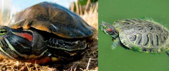
Pathological causes of soft shell
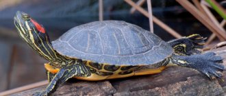
The dorsal shield of the shell in babies is bright green. With age, its color becomes darker, olive green. You can see black and yellow stripes on it. The ventral shield is dark in color and has black spots on it. These age spots on the red-eared slider's shell are a variant of the norm. At the same time, the shell is hard to the touch.
When it becomes soft and its separation is observed in an adult reptile, this is a symptom of pathology.
Often, a reptile has not only a soft shell, but a number of other signs:
- redness and swelling of the eyes;
- heat;
- the edges of the shell bend, its particles begin to peel off.
Such signs can be observed in various pathologies: dysfunction of the thyroid gland and intestines. The hardness of the shell depends on the amount of calcium in the reptile's body. The lack of this mineral causes the pyramidality of the carapace and a number of other signs and the development of rickets. The structure of the skull bones is disrupted, as a result the reptile cannot eat.
A lack of calcium in the body can be caused by dysfunction of the intestines and kidneys, which causes a deterioration in the absorption of the mineral. A deficiency of UV rays leads to a lack of vitamin D, which can also cause softness and peeling of the carapace tissue.
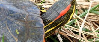
If your pet's shell peels off, this may be a sign of mycosis. With a fungal infection, a white coating may be observed on the shell of the red-eared turtle. White stripes can also remain on objects that the reptile rubs against. Also, when there is a fungal infection, the red-eared turtle scratches its shell.
In addition, white spots on the shell of a red-eared turtle can be observed if:
- the water in the aquarium is hard;
- improper feeding of the turtle;
- shell injuries;
- incorrect lighting.
Soft shells, bruises and an unpleasant odor can be observed due to burns and trauma to the shell, which are complicated by a secondary bacterial infection. In this case, the pet is treated with antibiotics.
If the reptile looks sick, you need to show it to a herpetologist as soon as possible, who will determine why the shell of the red-eared turtle is peeling off, it has become soft and will tell you what to do and how to help the animal.
How can you tell if a turtle is infected with a fungus?
It is extremely difficult for a non-professional to make an accurate diagnosis based on external manifestations alone. Although an experienced doctor can draw conclusions about the animal’s condition during an examination. But to identify a fungal infection, additional research is necessary. And after receiving the diagnostic results, the specialist will be able to tell whether your pet is suffering from an infectious disease.
It is extremely important to find the true reasons for the appearance of white plaque, and not act at random. Without an accurate diagnosis, you should not experiment with any drugs, especially antifungal agents, which can be very toxic and even life-threatening for the turtle. The use of certain products may cause the shell to peel off.
Diagnostic methods
First of all, the specialist examines the patient’s scalp, assessing the extent of the lesion and the appearance of dermatomycosis. To accurately determine the pathogen, the patient is prescribed laboratory tests, collection of material for bacteriological culture, microstudy, and also PCR (polymerase chain reaction). In addition, the patient is referred for blood and urine tests to determine his general health.
Microscopic diagnostic method
This method involves taking a scraping from the scalp or hair, which is examined under a microscope. Based on the appearance of the spores, a specialist determines the type of dermatomycosis. The internal structure of the hair also contains a certain type of fungal bacteria. Microscopic examinations are carried out from 3 to 5 days.
Microscopy is one of the methods for diagnosing scalp fungus
Scrapings and broken hair are also examined under a Voodoo lamp. In its ultraviolet glow, colonies of fungi and the affected area are clearly visible. Diseased areas will appear as a bright, unevenly outlined spot with a greenish-blue glow.
Bacteriological diagnostics
During bacterial sowing, material is taken from the area affected by the fungus and placed in a special instrument - a Peter's cup, with a medium previously diluted in it that is favorable for the proliferation of spores. The bowl filled with bacteria is placed in a thermostat for a certain time. Usually this is a period of two weeks. After growing the colony, the doctor determines the type and sensitivity of the pathogen to certain antifungal drugs.
PCR (polymerase chain reaction)
It differs from other research methods by studying biomaterial at the level of DNA and RNA after exposure to enzymes. This is the most modern and fastest way to obtain the necessary information about the nature of dermatomycosis and prescribe the correct treatment.
Prevention
It is almost impossible to eliminate contact with a parent suffering from a fungal disease, so it is necessary to cure the disease before the baby is born.
Until your loved one gets rid of the infection, you should minimize the risk of transmitting it to your baby: boil and iron bed linen, treat shoes with Formidron or vinegar essence, clean carpeting, disinfect the bathtub and bathroom floor.
When visiting public places with your baby where there is a risk of infection: the beach, swimming pool, bathhouse, put on rubber slippers. After the procedures, thoroughly dry your feet and wipe your feet with disinfectant solutions.
Scissors for cutting nails for a child must be individual.
You need to get rid of a contagious disease immediately and cure it until complete recovery. If a small child complains of itching, do not brush it off; the consequences of the infection can become apparent much later and can be very severe.
Fungus treatment
What is most interesting is that in natural conditions turtles practically do not get sick from the fungus. Some experts believe that the reason for this is captivity, in which basic instincts may be dulled, as well as a decrease in the effectiveness of the immune system.
In addition, a fungus in a pet can develop against the background of diseases such as pneumonia, tympany, as well as other negative factors that reduce the resistance of the immune system. Plus, this can be caused by the conditions of detention, as well as a meager diet. A lack of various vitamins, as well as inconsistency in temperature conditions, can serve as the trigger that will give rise to the development of infectious diseases, including fungus.
Most experienced owners can predict the appearance of fungus in advance, especially during periods after recovery from acute respiratory infections or other ailments that had to be treated with antibiotics. In this case, they prepare in advance to prevent the occurrence of mycosis. If you do not carry out such activities, the disease will soon remind itself and then it will be very difficult to cope with it.
The following types of fungi should be noted:
- Aspergillus spp;
- Candida spp;
- Fusarium incornatum;
- Mucor spp;
- Panicillium spp;
- Paecilomyces lilacinus.
In addition, at elevated water temperatures, the turtle spends a lot of time in the water without crawling onto land to warm up and dry out. Therefore, the temperature of the water should be maintained at 22°C-26°C, and the temperature of the island 28°C-32°C. Then the turtle will simply be drawn to the island to receive a certain dose of heat. Here it will dry out quickly and then the fungus is unlikely to be able to develop.
When arranging the habitat of the red-eared turtle, one should take into account such a thing as sufficient space for both living and rest. There should be enough space on the island for the turtle to feel comfortable and have the desire to constantly climb to this island. To make it convenient for her to do this, you should organize a smooth rise from the water.
It is quite easy to rid a turtle of a whole “bouquet” of infectious diseases if you follow the appropriate recommendations.
- Since there are several types of microbiotics for fungal infections, only a specialist can determine an accurate diagnosis. To do this, you will have to take a blood test and a smear. Only based on the test results can a conclusion be made about the type of fungal disease.
- Regardless of what kind of fungus the turtle has, it is necessary to completely disinfect the aquaterrarium, as well as the objects inside it.
- If several individuals are kept, the sick turtle is removed until the disease disappears. The turtle can be cured in two weeks or 2 months, depending on the severity of the disease.
- You need to add Methylene Blue to the water. Its quantity is selected so that the water is slightly colored and has a slightly bluish tint. If the aquaterrarium has a carbon filter, then it is better to turn it off, otherwise the effect of the drug will be neutralized.
- A sick turtle undergoes preventive and therapeutic actions. To do this, it is placed in a bath with oak bark tincture for 1 hour. This procedure is carried out daily until the fungus completely disappears. The color of the liquid should match the shade of regular tea. In severe cases, the concentration of oak bark can be increased.
- The turtle should be irradiated daily with a UV lamp. This radiation (ultraviolet) can destroy the infection.
- In addition to the above measures, the use of ointments is required. Ointments such as Triderm, Terbinofin, Akriderm and Nizoral are suitable for this. The turtle is smeared overnight, leaving it in a dry, warm place.
- In parallel with the treatment procedures, the sick turtle is prescribed a diet rich in vitamins and nutrients.
As a rule, such instructions refer to the main treatment regimen, although treatment with other methods is also possible. In any case, it is better to consult a veterinarian and not start self-treatment, which can lead to unpredictable consequences.
Disease prevention
There is one true truth: it is better to prevent a disease than to treat it. Owners with considerable experience do just this and regularly carry out preventive measures, especially when the turtle has an acute respiratory infection and a disorder in its digestive organs. When the body is severely weakened, all energy goes to fight the above ailments.
Preventive measures are carried out with the animal once every few months; for this purpose, the terrarium is etched with methylene blue. Having carried out prevention, you then need to provide the animal with normal conditions: ideal cleanliness, recommended temperature conditions and diet. All three components will help raise a healthy pet.
The turtle should receive ultraviolet irradiation for half of the day; for this, an appropriate lamp should be placed near it. There are special water conditioners; they need to be added to terrariums for preventive purposes. There are remedies against pathogenic microflora that can destroy infectious agents in a few days. For the drugs to have an effective effect, the terrarium must be in the shade. The ultraviolet lamp can only be turned on after a few days.
Parasitic fungi develop only on animals of the aquatic world and do not pose any danger to humans. When keeping several individuals in an aquarium, an animal with signs of disease should be quickly removed to prevent the infection from spreading to a neighbor. The fungus quickly spreads from one turtle to another.
Sometimes the cause of white crusty deposits is hard water. Lemon juice will help clean the shell if you wipe the stains with it several times. Such procedures should not be abused; it is better if there is constantly soft water in the aquarium; you can add special softeners to it. So, if there is a red-eared turtle in the house, the owner is responsible for its health. Organization of proper maintenance, balanced feeding and periodic services from veterinarians - only under such conditions this interesting animal will be able to delight with its presence for many decades.
Other interesting articles
- How much water does a red-eared turtle need in an aquarium? This species of turtle belongs to the family of freshwater turtles. In nature they can be found in small…
- Red-eared slider who can live with it Often, owners do not think about finding special equipment because they are going to keep the red-eared slider in...
- Can a red-eared turtle lay eggs in an aquarium? The simultaneous keeping of different-sex individuals of red-eared turtles at home, provided that optimal conditions are created, can...
How to treat fungus
Treatment of fungus in children requires an individual approach. The specialist must select the most safe medications for health
In this case, you need to pay attention to their effectiveness in the fight against mycosis.
If necessary, the doctor will recommend hospitalization for the child. This measure is usually required if the baby has concomitant problems that complicate the course of the fungal infection.
Drug treatment
Fungus of the nails on the hands and feet, as well as mycosis of the skin in children, is treated with medications. Complex or monotherapy is selected for the baby. In the first case, it consists of different groups of medications:
- Antihistamines;
- Immunostimulants;
- Vitamin complexes;
- Hormonal drugs;
- Antifungal agents of local and systemic action.
During treatment, it is recommended to use tablets and ointments to increase the effectiveness of the therapeutic course. For children with an infection, for example, the little finger or the oral mucosa, the following medications are suitable:
- "Miconazole";
- "Limisil";
- "Exoderil";
- "Clotrimazole".
Antifungal ointments are recommended to be applied to the skin and nail plates about 2-3 times a day. It is best to treat the infected areas with topical medications in the mornings and evenings. This course of treatment is usually followed for 1.5 months.
If nail fungus in children continues to progress, then treatment is supplemented with tablets. They fight pathogenic microflora from inside the body.
Systemic therapy for fungus in children is usually carried out using the following means:
- "Diflucan";
- "Griseofulvin";
- "Terbinafine".
Ointments containing zinc, tar or salicylic acid also help in the fight against lichen in a child. In particularly severe situations, the use of hormonal drugs is required.
Yeast fungus in children that is found in an intimate place is recommended to be treated with Mycelex and Monistat suppositories.
It is prohibited to independently treat mycosis in a child.
Folk remedies
Not only traditional, but also traditional medicine helps cure a nail or skin from mycosis. The methods she proposed are safe for the child. Fungus of the nail, mucous membrane or skin can be treated with the following means:
- Celandine. 20 g of plant material should be poured into 1 liter of hot water. When the composition is infused, it needs to be warmed up a little, and then the limbs affected by the fungus are dipped into the infusion. If the infection has affected other parts of the body, they should be wiped with a cotton pad soaked in this product;
- Natural honey. Helps with mycosis of the skin in children. The product in an amount of 1 liter must be mixed with 10 liters of water. This solution is recommended for use for preparing lotions;
- Phytotherapy. A good result can be achieved if you use sage, oak bark, St. John's wort and chamomile in the fight against fungus. Herbs must be used separately. Medicinal infusions are prepared from them, which are suitable for lotions and baths. To make the medicine, just pour 20 g of herb into 200 ml of boiling water and leave for 15 minutes;
- Sea salt. It is recommended to steam limbs affected by the fungus in a solution based on it. To prepare the medicinal composition, only 4-5 tbsp is required. l. salt;
- Mint. Suitable for treating feet infected with fungus. 50 g of fresh mint leaves should be poured into 2 identical bags. Afterwards you need to put them on your feet and secure them with socks. It is recommended to wear peculiar compresses for 2 hours;
- Tea tree oil. You need to moisten cotton pads in it and use them to treat infected areas of the child’s body.
All questions regarding the use of folk remedies in the treatment of a child must be discussed with the attending physician.
Diet
Toenail fungus in children, as well as on other parts of the body, can be overcome with a special diet. It is part of the treatment course. When compiling a child’s diet, it is recommended to include vegetables, fruits, seeds, chicken eggs and dairy products in his menu. Your baby can drink unsweetened tea.
Heavy and unhealthy food will only harm your child. Therefore, it is advisable to abandon it
It is especially important to avoid consuming dairy products, yeast breads and sweets, as they can provide food for pathogenic microorganisms
The fungus “loves” sweets, so during treatment you need to avoid baking and sweets
Features of care during molting
During the molting period, the reptile needs special care. She needs to continue to be fed as before, but the pet’s diet will need to be changed. The animal requires more calcium and vitamins.
During molting, it is worth giving your pet shrimp and lean fish, for example, hake. To replenish the reptile’s body with calcium, they must be fed along with the bones. There is no need to be afraid that your pet will choke; its jaws are designed for chewing such food.
After consultation with a herpetologist, you can give special multivitamins designed specifically for young animals. During the molting period, it is important to monitor the cleanliness of the water in the aquaterrarium where the reptile lives. After all, at this time you can notice pieces of skin and shell on the surface of the water, which can begin to rot and cause the pet to become ill. To prevent this, you need to change the water in the terrarium as often as possible
Sometimes owners try to help the reptile get rid of flaking skin and shell. But this is allowed only in extreme cases, if the dead tissue does not fall off for a long time and causes discomfort to the animal. You can wipe the turtle with a decoction of chamomile flowers or use a soft toothbrush to clean the animal’s shell. This must be done carefully so as not to injure the reptile.
If the owners do not pay attention to the exfoliated areas of skin for a long time, the pet may get sick or scratch itself, trying to get rid of the dead epithelium. You can take the reptile out of the aquaterrarium for a while and wait until the animal is completely dry. Dry tissue will peel off more easily and fall off when your pet swims in the water.
During molting, experts recommend bathing the animal. This is necessary to remove remaining dirt from under the shell scutes and to prevent them from rotting. To bathe your pet, you need a shallow dish with low sides. Pour warm water into it and add baking soda. You need 7 g per liter of water
Carefully lower your pet into the liquid. In this case, you need to ensure that the reptile’s head remains on the surface.
The duration of the bath is 20 minutes. After its completion, excess water is blotted with a towel, and the shell is greased with olive oil. Just a few drops are enough.