Description and difference from fungal infection
Columnaria affects the body of the fish, so the disease is often confused with a fungal infection. This leads to an incorrectly selected treatment regimen and the death of animals. Columnaria can be distinguished by the characteristic signs of the disease. With columnaria, the formations on the body are gray in color, they are rough and large. The disease does not develop quickly, it manifests itself gradually. A characteristic feature is white fluff near the mouth, fins, and scales.
The disease progresses in fresh water at high water temperatures of 28-30 degrees. The bacteria affect fish with weakened immune systems. Infected fish become dangerous to other inhabitants of the aquarium. When bacteria enters an aquarium among fish, the infection spreads quickly, so it is important to remove the sick fish from the common tank to prevent further infection.
The disease can occur with characteristic symptoms or asymptomatic. The fish dies 2-3 days after infection. If the spread of bacteria is not noticed in a timely manner, all inhabitants of the reservoir may die.
Most often, the disease affects labyrinthine and viviparous species.
Flexibacteriosis Clinic
Bacteria affect bone cartilaginous tissue, gills, and internal organs. Almost always, the disease affects all of the listed systems, however, according to the predominance of certain symptoms, it is possible to distinguish variants of the clinical course of the disease. According to my observations they are as follows.
- "Mouth fungus." This option is most often described in the literature. It initially manifests itself in the formation of plaque around the fish’s mouth. The color of the plaque can vary from a barely noticeable light gray to bright white, I would say fluorescent, as if the fish had white paint on its lips. The visibility of this plaque also depends on the color of the fish itself; it is hardly noticeable, and therefore the disease is diagnosed when the next stage begins - the destruction of osteochondral tissue. Bacteria secrete enzymes that destroy cartilage, thereby creating a nutrient medium for themselves, so the next stage is the occurrence of deformations in the “face” of the fish. The deformation begins from the mouth opening - the mouth does not close, a “leaky mouth opening”, sometimes the mouth opening moves to the side. The antennae of catfish “dissolve.” If the course of the disease is interrupted due to treatment or self-healing (and this is possible), then deformations of the skull remain for the rest of life. If the deformations are significant, then the fish cannot feed normally and eventually dies.
- "Gill" form. The gills are always affected by columnar blight, but the clinical picture is influenced by the degree of their damage. In the form of the disease with predominant damage to the gills, symptoms of oxygen starvation come to the fore. At first, the fish becomes lethargic, the movement of the gill covers becomes more frequent, later the fish stays near the surface, and when swimming it begins to “sway”. In case of severe damage, the fish stands at the surface, small fish literally lie down on a floating Riccia bush (by the way, they are often found dead there). Severe symptoms indicating imminent death are the absence or extremely weak reaction to external stimuli, as well as a clearly visible symptom that I have observed more than once - standing at the surface, the fish seems to arch its back, trying to stick its mouth out of the water.
- “Pseudohydropsea” form. Indicates damage to internal organs. The main symptom is a swollen abdomen. Unlike the “classic” one, there is no bulging eyes or ruffling of scales. The combination of these two symptoms most likely indicates a mixed infection. In addition to abdominal bloating, there are a number of characteristic symptoms. The first thing that catches your eye is the compression of the fins; the fins themselves are most often unchanged. Swimming movements become strange, worm-like. The second symptom is constipation with the release of thin, transparent excrement. I do not undertake to definitively judge whether this symptom belongs to columnaria itself or to concomitant hescamitis (spironucleosis). I am inclined to the first point of view, since therapeutic measures aimed at flagellates do not give a clear effect, and antibacterial therapy allows for improvement.
- "Pseudotuberculous" form. Unlike the previous form, here, on the contrary, there is significant emaciation. This course of the disease is typical for a protracted variant (1 in 1). Weight loss develops gradually. The fish becomes shy, refuses to take food and gradually loses weight; the sunken abdomen is especially noticeable, which is why the fish can take on a “humpbacked” appearance. It flows slowly, but almost always leads to death.
Associated symptoms include separation, erosion of the fins, and the appearance of plaque on the fins. Sometimes the “oral fungus” is localized on the fins - then marginal erosion with clearly defined edges, covered with a white or gray coating, forms on the fin. Without treatment, the fin may completely collapse. In practice, I have seen such destruction of the tail fin (in catfish, small tetras). In severe cases, the process does not stop there - ulceration of the soft tissue occurs. The ulcer is usually surrounded by a gray or white coating. Such a fish dies quickly. All forms of columnarosis can be observed simultaneously, but one of them usually predominates. The most malignant, according to my observations, is the gill form. Pseudohydropseal and pseudotuberculosis forms are treated rather poorly, and the form of “oral fungus”, although treated well, leaves behind deformation of the skull bones. I will describe a couple of cases of the disease with different courses, then a few words about diagnosis as such and treatment.
An acute form of the disease arose in a completely healthy aquarium. First, dead male handsome guppies were noticed on the Riccia bushes - without any visible abnormalities, only dull coloring. This was the reason for the consultation. An aquarium with good conditions, no overcrowding, no stress, T 24-25, water parameters are normal. The disease developed after the purchase and release of 2 female guppies without quarantine (after about 5-7 days). The population of the aquarium includes livebearers and catfish. The aquarium contained fish of different ages - juveniles grew right in the aquarium (platies, guppies). Upon examination, more than half of the population is affected - adult fish with a bright white coating on the mouth (not all), purchased females with a swollen abdomen without ruffling of scales, fins are compressed, kept near the surface, and make worm-like movements when swimming. Young fish (not all) have a swollen abdomen and compressed fins. 3 fish near the surface, greedily swallowing air. A juvenile platy has half his caudal fin eroded. Approximately half of the population does not show obvious symptoms of the disease. This is an acute course of columnarosis, which has taken on the character of an epidemic. It can be predicted that after a few days the disease will spread to all those remaining. It is clear that the disease manifested itself in different individuals with a predominance of different symptoms.
With a slow, protracted flow, the picture is somewhat different. Also a relatively healthy aquarium. Over the past 2 months, fish (Neons and livebearers) began to die. 1-2 deaths per month. In this case, no external changes other than a swollen or slightly sunken abdomen are observed. The reason for the consultation was “Pecilias are losing weight.” Upon examination, large female platies have a sunken abdomen; the fish are shy, hide in plants, eat, but little by little. Water parameters - high level of nitrates, some overcrowding of the population. Planting in an aquarium through quarantine. I carefully study the population of the aquarium - nothing. The second time I was present at the feeding - I noticed that one minor was taking food in a strange way - deformation of the jaws - and then I noticed a barely earthy gray coating around the mouth, some fraying of the fins. Without looking closely, you won’t notice such a plaque; if not for the deformities, the disease would remain undiagnosed.
Columnaria: causes
The main reasons for the appearance and spread of bacteria in an aquarium are overcrowding of the tank and a small area. The progression of the disease is promoted by:
- high organic content;
- lack of oxygen;
- high level of acidity;
- high concentration of ammonia substances, nitrites;
- external injuries in fish;
- heat.
Bacteria can enter an aquarium with infected fish from a pet store, so it is important to keep new inhabitants in quarantine for the first weeks.
Protracted cases of flexibacteriosis
As always, in cases of indolent diseases, it is necessary to establish optimal conditions for keeping fish. It has been noticed, for example, that charantsinus get sick much more often at a pH of 8 than at a pH of 6.5. feeding corresponds to the needs of the fish - for example, green food is required for livebearers. OPTIMAL CONDITIONS ARE YOUR ALLY IN THE FIGHT OF DISEASE. If, despite the creation of ideal conditions, the disease periodically sends one fish or another to the next world, fish with suspected disease should be isolated and treated in a hatchery with doxycycline. The rest of the fish can be fed with medicated food (I used Bacto Tabs). It is almost impossible to cure fish with advanced stages of the disease (for example, severe emaciation). But manifestations of “oral fungus” are treated, but leave behind bone deformations (I have one of these that has been living for 2 years now and is not going to die). We pass the water in the general aquarium through a UV sterilizer (3-5 days). If there are no sensitive plants in the aquarium and only livebearers live, you can add a little salt to the water. Otherwise, after sterilizing the water, I added methylene blue until the water turned slightly blue. Contrary to what is written, in such doses the blue never disrupted biofiltration. At the end of such 3-4 days of therapy, the bluing can be easily removed by substitutions or a carbon filter. If, despite this, a relapse of the disease occurs after some time, then therapy is carried out in a general aquarium according to the scheme for acute cases.
Forum thread
No entries found.
Symptoms
Columnaria can be recognized by its characteristic symptoms, but you need to be prepared that in some cases the disease is asymptomatic:
- rocking the fish in place;
- moving the fish jerkily;
- loss of interest in what is happening;
- pinching fins;
- gray spots near the mouth, on the gills, fins, head;
- the appearance of white fluff near the oral cavity, on the body;
- coloring the scales white;
- the appearance of bloody smudges on the body;
- decomposition of gills;
- change in color of gills in fish;
- bulging eyes;
- fin decomposition.
The disease spreads quickly, so it is important to start treatment of the sick individual in a timely manner. White spots grow rapidly, spreading throughout the body. Columnaris bacteria eat away the skin down to the muscles, which leads to the death of pets within 2-3 days.
Pathogen
Flexibacter columnaris is a rod-shaped bacterium that grows optimally at a temperature of 28-30°C. Moves by sliding. High temperatures stimulate the growth of these bacteria. Although the bacteria still continues to grow at 4°C, even at 12°C there is no infection. Growth stops in salt water, so these bacteria mainly affect freshwater fish. This bacterium is often found in our aquariums and affects heat-loving fish, such as tropical ornamental fish.
Columnaria: treatment
Treatment of columnarosis should not be delayed, as the disease spreads quickly, affecting all weakened fish. If symptoms are detected in one fish, you need to be prepared that other infected fish will appear, so you need to treat all the inhabitants of the aquarium at once.
The first step is to reduce the acidity level of the water to 6 pH. Acidity is reduced gradually so as not to cause osmotic shock in the inhabitants.
Levomycetin is dissolved in a general aquarium. The antibacterial agent quickly kills bacteria, reducing the risk of infection spreading. Levomycetin is added to the tank, maintaining a proportion of 4 grams of the drug per 100 liters of water. During treatment, it is necessary to maintain a constant temperature in the aquarium no higher than 27 degrees. Treatment with chloramphenicol is carried out for 5 days, with daily water changes. If the water in the tank becomes cloudy, it is recommended to replace the water by a quarter to reduce the concentration of chloramphenicol. To enhance the effect of the drug, add 4-5 tablespoons of salt per 100 liters to the water.
Separately, baths with phenoxyethanol are prepared for sick fish. The fish are placed in the bath for a while and then removed to a common tank. This treatment brings quick, positive results. Instead of phenoxyethanol, the drug Merbromin, methylene blue, is often used.
When the disease penetrates the internal tissues, strong antibiotics are used. They are bought in specialized stores or pharmacies.
After treating aquarium fish, the tank is completely disinfected.
Preventive actions
Columnaria is a common disease that is difficult to cope with. Therefore, experienced aquarists recommend taking preventive measures. After all, a disease is easier to prevent than to treat.
- Selection of an aquarium. For each phenotype, a reservoir with the required parameters and characteristics is selected. Some phenotypes require containers that are quite large in volume. Each type of fish requires an appropriate volume.
- Condition of aquarium water. Remains of organic substances are removed from aquarium water. Remains of nutrients are removed from the bottom. To facilitate the process, experienced aquarists select the appropriate substrate. In addition, the soil is regularly siphoned using appropriate equipment for this purpose.
- Columnaria does not develop in water that is saturated with active oxygen. An aquarium with fish is equipped with an oxidizer or other unit with which the water is saturated with oxygen. Timely introduction of the setting helps reduce the likelihood of developing the disease.
- Quarantine is carried out periodically. Salted liquid is poured into a container designed for this purpose. This event is carried out provided that the phenotype normally tolerates the presence of salt.
- Columnaria practically does not develop in acidic and soft water. Therefore, these indicators are maintained within the established limits. Modern units are used to monitor acidity and hardness.
- Water changes are performed 1–2 times during the day. Experienced aquarists carry out this procedure until all symptoms are eliminated.
- Filter. The tanks are equipped with mechanical or chemical filters. They are cleaned every 7–14 days. Purified aquarium water is used for washing.
Quick identification of columnaria and proper treatment allows you to protect all phenotypes that are present in the aquarium. Beginners and experienced aquarists prepare first aid kits in advance in which they place medications. Before carrying out events, consultation with specialists is required.
Video about diseases of aquarium fish
AdminAuthor of the article
Did you like the article?
Share with your friends:
Prevention
To prevent your fish from becoming infected with Columnaria, you must be attentive to the new inhabitants and maintain hygiene and cleanliness of the aquarium.
Before purchasing, you need to carefully examine the fish in a store aquarium. If there are fish in the tank with signs of columnaria, you should postpone the purchase or select fish from another container.
New fish cannot be immediately transplanted into a common aquarium; they are kept in quarantine for 1-2 weeks, monitoring their condition. If the fish is pinching its fins or withdrawing into seclusion, it may be sick.
You should not overpopulate the aquarium. It is necessary to maintain the proportion so that there is at least 1 liter of water for each fish. In crowded spaces, the likelihood of bacteria spreading increases several times.
The pond should be cleaned regularly. Preventive measures include changing the water, cleaning the walls, and siphoning the soil.
Columnaria disease can become a real disaster for a home aquarium. This disease can quickly destroy all abodes, so it is important to monitor the aquarium, temperature, and cleanliness. If the inhabitants of a reservoir develop symptoms of columnaria, treatment should be started immediately. The water in an infected aquarium is changed daily until signs of the disease disappear.
About diagnostics
“Mouth fungus” (and this name is incorrect) must be distinguished primarily from saprolegnia. Anyone who has seen both is unlikely to confuse them. Saprolegnia is essentially a mold and forms a coating in the form of cotton wool or a “bush of mold” where threads are visible. The coating of columnaria is uniform, velvety, or even glossy. It differs from parasitic infestations, which are also accompanied by a visible mucous coating or discoloration, primarily by localization - the mouth and fins. Apparently, infection with Columnaria does not cause itching, and I have never noticed that the fish itch, while with infestations, “scabies” is a common occurrence. The gill form is quite difficult to distinguish from other conditions accompanied by damage to the gill filaments. Noteworthy is the rate of increase in symptoms (with columnarium it progresses quickly), the epidemic nature of the infection (there may be fish in the aquarium with other forms of the disease), and again the lack of scratching. Pseudodropsy must be distinguished from physiological conditions (“pregnancy”, viviparity, obesity), or from a number of pathologies (true dropsy). The pseudotuberculosis form must be distinguished from tuberculosis and diseases caused by sporozoans (for example, plastophora).
I was "lucky". I have encountered this disease more than once both in my aquarium and among friends. Being an amateur aquarist, I do not conduct serious ichthyopathological studies; everything written is the result of observations, studying various literature (including foreign sources that have not been translated here), as well as some hypotheses that I am trying to formulate, having some experience. Columnaria is a complex disease. Despite the fact that this is a bacterial disease, it is always considered apart from other bacterial infections. To put it bluntly, in a significant number of cases it is closer in manifestations to tuberculosis than to a “bacterium”. The causative agent of the disease is a bacterium. It had and still has many names, but at the moment it is considered correct to call it Flavobacterium columnare. This is a gram-negative rod that can grow even in the presence of some antibiotics (for example, neomycin and polymyxin). Produces enzymes that destroy chondroitin sulfate, one of the components of cartilage tissue. The disease occurs frequently, probably much more often than we think. The problem is that it is often difficult to diagnose. There may be no obvious symptoms at all; in quarantine the disease is not recognized and therefore the bacterium easily enters a common aquarium, where it finds quite favorable conditions for itself, causing epidemics. In addition, the already developed symptoms of the disease are such that they require close analysis and differentiation from other similar diseases. According to some foreign sources, there are four types of disease according to their severity and speed. With the most severe - lightning variant, the fish die within 48 hours, and with the most erased protracted (which I nicknamed 1 in 1), the disease quietly takes the lives of 1-2 fish per month. It is not entirely clear whether these variants are caused by different types of bacteria, or whether it all depends on the conditions, but most authors are inclined to the first point of view. Many authors call columnaria blight a “fish stress disease.” This means that the disease develops against the background of acute or chronic stress, being, as it were, an indicator of it. Such conditions, for example, are often created when feeding a large number of young fish. And one more small but important note. The Columnaria bacterium is heat-loving; its optimal growth temperature is about 27-28 degrees. And she absolutely cannot tolerate salt (as the famous line from the film says, “We need to take advantage of this.”)CLINIC Columnaria bacteria affect osteochondral tissue, gills, internal organs; the focus of the disease can also be localized on the external integument; in advanced cases, erosion occurs, and then ulceration, involving the underlying muscle tissue. Almost always, the disease affects all of the listed systems, however, according to the predominance of certain symptoms, it is possible to distinguish variants of the clinical course of the disease.
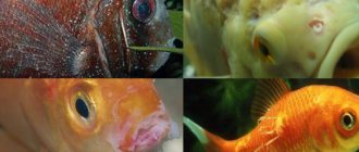
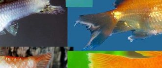
Photo 1. The photo shows a young barb affected by Columnaria.
A characteristic plaque is visible in the area of the pectoral fin, spreading to the head. In the area of plaque, a depression (erosion) is observed, which is very characteristic of columnaria. The plaque can also be seen on the muzzle, more clearly on the lower jaw. This individual begins to develop deformations of the skull bones. First of all, it must be said that there are external (more well known to us from the literature) and internal forms of the disease. Data on the frequency of various forms do not coincide among different authors, but on average 20% of all cases are internal forms, 50% are mixed, and 30 are external. Internal forms occur with damage to the internal organs - while we do not see any plaque, cloudiness, or “dissolution” of the fins (or these symptoms exist, but are expressed so little that we do not notice them), but the fish loses weight, compresses the fins, and begins to swim strangely . If we try to somehow systematize the symptoms of the disease into groups, we will get the following options (forms of the disease). 1. “Mouth fungus.” This option is most often described in the literature. It initially manifests itself as the formation of plaque around the fish’s mouth. The color of the plaque can vary from a barely noticeable light gray to bright white, I would say fluorescent, as if the fish had white paint on its lips. The visibility of this plaque also depends on the color of the fish itself; it is hardly noticeable, and therefore the disease is diagnosed when the next stage begins - the destruction of osteochondral tissue. Bacteria secrete enzymes that destroy cartilage, thereby creating a nutrient medium for themselves, which is why deformation of the “face” of the fish occurs. The deformation begins from the mouth opening - the mouth does not close, a “leaky mouth opening”, sometimes the mouth opening moves to the side. The antennae of catfish “dissolve.” If the course of the disease is interrupted due to treatment or self-healing (and this is possible), then deformations of the skull remain for the rest of life. If the deformations are significant, then the fish cannot feed normally and eventually dies. I also include “irregular oral fungus” in this form - this is the option when the plaque is localized not in the mouth, but on any other part of the body. 2. “Gill” form. The gills are always affected by columnar blight, but the clinical picture is influenced by the degree of their damage. In the form of the disease with predominant damage to the gills, symptoms of oxygen starvation come to the fore. At first, the fish becomes lethargic, the movement of the gill covers becomes more frequent, they can protrude, later the fish stays near the surface, and when swimming it begins to “sway”. In case of severe damage, the fish stands at the surface, small fish literally lie down on a floating Riccia bush (by the way, they are often found dead there). Severe symptoms indicating imminent death are the absence or extremely weak reaction to external stimuli, as well as a clearly visible symptom that I have observed more than once - standing at the surface, the fish seems to arch its back, trying to stick its mouth out of the water. After the death of the fish, the same plaque can be found on the gill filaments as the “mouth fungus”. 3. “Pseudohydropsea” form. Indicates damage to internal organs. The main symptom is a swollen abdomen. Unlike the “classic” one, there is no bulging eyes or ruffling of scales. The addition of these two symptoms most likely indicates a mixed infection, that is, the “connection” of a new pathogen to the disease. In addition to abdominal bloating, there are a number of characteristic symptoms. The first thing that catches your eye is the compression of the fins; the fins themselves are most often unchanged. Swimming movements become strange, worm-like. The second symptom is constipation with the passage of thin, clear excrement. I do not presume to definitively judge whether this symptom belongs to columnaria itself or to the concomitant hescamitosis (spironucleosis) - although the causative agent of columnaria tends to create associations, that is, “make friends” with other pathogens. I am inclined to the first point of view, since therapeutic measures aimed at flagellates do not give a clear effect, and antibacterial therapy allows for improvement. 4. “Pseudotuberculosis” form. Unlike the previous form, here, on the contrary, there is significant emaciation. This course of the disease is typical for a protracted variant (1 in 1). Weight loss develops gradually. The fish becomes shy, refuses to take food and gradually loses weight; the sunken abdomen is especially noticeable, which is why the fish can take on a “humpbacked” appearance. It flows slowly, but almost always leads to death. Associated symptoms include delamination, erosion of the fins, and the appearance of plaque on the fins. Sometimes the “oral fungus” is localized on the fins - then marginal erosion forms on the fin with clearly defined (as if a piece had been bitten off) edges covered with a white or gray coating. Without treatment, the fin may completely collapse. In practice, I have seen such destruction of the caudal fin (in catfish, small tetras). In severe cases, the process does not stop there - ulceration of the soft tissue occurs. The ulcer is usually surrounded by a gray or white coating. Such a fish dies quickly. All forms can be observed simultaneously, but one of them usually predominates. The most malignant, according to my observations, is the gill form. Pseudohydropseal and pseudotuberculosis forms are treated rather poorly (you need to be able to “catch them” in time - and this is very difficult), and the form of “oral fungus”, although treated well, leaves behind deformation of the skull bones.
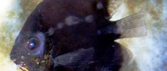
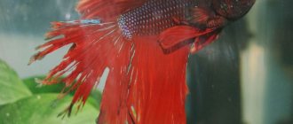
Photo 2. Characteristic erosion of the fin due to columnar blight. A white coating is clearly visible along the edge of the erosion. The edges are crisp.
I will describe a couple of cases of the disease with different courses, then a few words about diagnosis as such and treatment. 1. An acute form of the disease arose in a completely healthy aquarium. First, dead male handsome guppies were noticed on the Riccia bushes - without any visible abnormalities, only dull coloring. This was the reason for the consultation. An aquarium with good conditions, no overcrowding, no stress, T 24-25, water parameters are normal. The disease developed after the purchase and release of 2 female guppies without quarantine (after about 5-7 days). The population of the aquarium includes livebearers and catfish (corydoras). The aquarium contained fish of different ages - juveniles grew right in the aquarium (platies, guppies). Upon examination, more than half of the population is affected - adult fish with a bright white coating on the mouth (not all), purchased females with a swollen abdomen without ruffling of scales, fins are compressed, kept near the surface, and make worm-like movements when swimming. Young fish (not all) have a swollen abdomen and compressed fins. 3 fish near the surface, greedily swallowing air. A juvenile platy has half his caudal fin eroded. Approximately half of the population does not show obvious symptoms of the disease. This is an acute course of columnarosis, which has taken on the character of an epidemic. It can be predicted that after a few days the disease will spread to all those remaining. It is clear that the disease manifested itself in different individuals with a predominance of different symptoms. But this epidemic nature of the disease forces us to look for a single pathogen. 2. With a slow, protracted flow, the picture is somewhat different. Also a relatively healthy aquarium. Over the past 2 months, fish (Neons and livebearers) began to die. 1-2 deaths per month. In this case, no external changes other than a swollen or slightly sunken abdomen are observed. The reason for the consultation was “Pecilias are losing weight.” Upon examination, large female platies have a sunken abdomen; the fish are shy, hide in plants, eat, but little by little. Water parameters in - high level of nitrates, some overcrowding of the population (and if we suspect Columnaria, especially sluggish, we always find out all the factors that can stress the fish). Planting in an aquarium through quarantine. I carefully study the population of the aquarium - nothing. The second time I was present at feeding - I noticed that one minor was taking food strangely - deformation of the jaws - and then I noticed a barely noticeable gray coating around the mouth, some fraying of the fins. Without looking closely, you won’t notice such a plaque; if not for the deformities, the disease would remain undiagnosed.
Now about diagnostics.
“Mouth fungus” (and this name is incorrect) must be distinguished primarily from saprolegnia. Anyone who has seen both is unlikely to confuse them. Saprolegnia is essentially a mold and forms a coating in the form of cotton wool or a “bush of mold” where threads are visible. The coating of columnaria is uniform, velvety, or even glossy. It differs from parasitic infestations, which are also accompanied by a visible mucous coating or discoloration, primarily by localization - the mouth and fins are a favorite place. Apparently, infection with Columnaria does not cause itching, and I have never noticed that the fish itch, while with infestations, “scabies” is a common occurrence. The gill form is quite difficult to distinguish from other conditions accompanied by damage to the gill filaments. Noteworthy is the rate of increase in symptoms (with columnarium it progresses quickly), the epidemic nature of the infection (there may be fish in the aquarium with other forms of the disease), and again the absence of scratching. Pseudodropsy must be distinguished from physiological conditions (“pregnancy”, viviparity, obesity, caviar in females), or from a number of pathologies (true dropsy). The pseudotuberculosis form must be distinguished from tuberculosis (but this is difficult to do, given that these two diseases can go “hand in hand”) and diseases caused by sporozoans (for example, plistophorasis). PREVENTION The question remains whether the causative agent of Columnaria is a conditional pathogen (that is, the disease develops only under some unfavorable conditions), or whether this bacterium will always cause disease even in minimal quantities. From my practical experience, I am more inclined to the second option. But there is one big BUT. What form of the disease are we talking about? about acute, subacute, sluggish, protracted? Can one form transform into another? Preliminary studies by foreign authors indicate that they cannot - these are different strains with different virulence and pathogenicity. Therefore, perhaps the truth lies somewhere in the middle - acute forms of the disease are contagious and spread like an epidemic, while protracted forms could be called a disease of containment and stress; when favorable conditions arise, they can subside. At the same time, the authors provide data on morbidity in nature and the following picture emerges - the aquarium itself is stressful. Be that as it may, columnarium should be considered a highly contagious disease. And the pathogen itself, even in water without a substrate (fish, plants, filter), persists for up to 45 days.
TREATMENT. There are many methods and treatment regimens described in various literature, ranging from table salt and methylene blue. and ending with combinations of 4-5 antibiotics. Let's break it all down. First, general comments, then acute cases, and then a protracted course. Columnaria is a systemic (that is, with simultaneous damage to many organs) bacterial disease in 80% of cases. For such diseases it is necessary to use antibiotics. The drugs of choice are antibiotics of the tetracycline group. Can it be treated with other antibiotics? Yes, it is possible (there are treatment regimens with kanamycin and nitrofurans), but the treatment effect will be much less reliable. Dyes and antiseptics, although they kill bacteria directly in the water and on the surface of the fish, are ineffective against bacteria that have penetrated into tissues and organs. In human pharmacies you can find 3 drugs (more in veterinary ones) - tetracycline, metacycline and doxycycline. I prefer the last two. Why? For therapy, the antibiotic must dissolve well in water and form stable solutions. Tetracycline hydrochloride seems to have these properties. I recently went to 10 pharmacies and none of them had hydrochloride. Tetracycline, the base that is proposed, is NOT suitable for aquarium therapy - very low solubility. Therefore, be careful when using pharmaceutical drugs. In addition, tetracycline in aqueous solutions is unstable and therefore is administered in shock dosages so that by the end of the day at least some concentration of the drug remains. Then the introduction is repeated. Such a wave-like fluctuation in the concentration of the drug in water is the best training for bacteria to form resistance (resistance) of bacteria to the action of the antibiotic. Since the treatment is long-term and repeated courses may be required, this should not be allowed. Therefore, when treating columnaria, I DO NOT use tetracycline. Metacycline and doxycycline are available in the form of hydrochloride in convenient capsules, which, when opened, yield a fine powder that instantly dissolves in water. The drugs are similar in spectrum of action to tetracycline, but have a number of valuable properties. Metacycline forms stable solutions in which the active substance is destroyed extremely slowly. For comparison, tetracycline disintegrates in solutions by 30% after 8 hours, and by another 30% after 24 hours. Doxycycline holds the record for penetration through biological membranes. There is no exact data, but apparently it penetrates through the gills, and the drug has an inherent property of accumulation due to its long period of decay in biological tissues. However, its aqueous solutions decompose in bright light, so you have to turn off the lighting. When treating with tetracycline antibiotics, remember the following: in hard water, their effectiveness is reduced due to the ability to bind and form insoluble compounds with calcium and magnesium ions. (dosage needs to be increased). And second, efficiency is maximum at pH values close to neutral. In acidic and alkaline environments, the decomposition of the active substance is accelerated. (pH range 6-8 can be considered “close to neutral”). THERAPY OF ACUTE CASES. Treatment is carried out in a common aquarium, since acute cases are always an epidemic. I use a combination of doxycycline and biseptol. The addition of biseptol makes it possible to reduce the dosage of both drugs by 2/3 due to their mutually reinforcing effect, which is more optimal from the point of view of toxicity. With this combined use, the dosage of doxycycline is 200 mg (2 capsules) per 100 liters, the dosage of Biseptol-480 is 1.5 tablets per 100 liters. If the water is hard, the dosage of doxycycline is increased by ¼. First, biseptol is added. The tablets are crushed into powder and poured into a glass of warm water. Mix vigorously. Let it sit for 30 minutes and then stir again. Undissolved particles will dissolve directly in the aquarium. No damage occurred when the particles hit the plants. Then the suspension prepared in this way is poured in a thin stream into the aquarium under a sprayer or into a filter stream. Undissolved particles in the glass dissolve in the aquarium within approximately 30-60 minutes. Then doxycycline is added. The powder from doxycycline capsules dissolves well even in a small (100-200 ml) amount of cold water, resulting in a clear, slightly yellowish solution. This mother liquor is then applied in a thin stream under a sprayer or filter. If fractional dosage is necessary (for example, half a capsule is required), do not divide the powder. Dissolve the whole capsule in 200 ml of water, pour 100 ml into the aquarium, and put the rest in the refrigerator. In a dark place the solution is stable for 2-3 days. The lights in the aquarium are turned off; if there is direct sunlight, they are covered with a curtain. When water is very hard, doxycycline complexes give the water a slightly brownish color. This usually becomes noticeable on day 2-3 of treatment. Antibiotic therapy is continued for 5 - 7 days. Sometimes it is necessary to increase the duration of treatment to 10 days. Every day we replace 25-30 percent of the water and add 100 mg per 100 liters (1 capsule of doxycycline). and 0.5 tablets (per 100 l) of Biseptol 480. I remind you that when treated with antibiotics in a general aquarium, biofiltration suffers (I won’t go into detail). And the second note is plants. To prevent them from completely bending, immediately before the daily water change I turn on the light for 2-3 hours. At the same time I feed the fish. With the gill form, the fish often die faster than the antibiotic can take effect. Here, I think, it makes sense to bathe such fish in a concentrated salt solution (5-10 grams per liter) before starting treatment. The purpose of this is the following: Columnaria bacteria do not tolerate mineralization, perhaps we will be able to stop the growth of colonies on the gills, and then the antibiotics will finish their job. And secondly, perhaps osmotic stress will prolong the life of the fish. I would like to emphasize that such baths are carried out with a gradual increase in salinity (as expected, according to all the rules), and specific techniques are described in the literature. It's worth trying anyway, but the prognosis is still unfavorable. From the third day I begin to administer drugs that accelerate tissue repair (restoration). For this purpose I use aqueous extract of propolis (1 teaspoon per 100 liters every other day). Some fish begin to itch when it is applied, but as a rule, this goes away after 30-40 minutes. There is evidence that treatment of external symptoms of the disease can be accelerated by administering pimafix in doses recommended by the manufacturer. In severe cases, it is permissible to increase the INITIAL dose of doxycycline by 1.5-2 times. The dose of Biseptol should not be increased. At the end of treatment (under normal conditions this is a 7-day course), it is important to quickly start biofiltration, or at least prevent the appearance of ammonia and nitrites in the water. PROTIGED CASES. As always, in cases of indolent diseases, it is necessary to establish optimal conditions for keeping fish. It has been noticed, for example, that charantsinus get sick much more often at a pH of 8 than at a pH of 6.5. feeding must meet the needs of the fish - for example, green food is required for livebearers. OPTIMAL CONDITIONS ARE YOUR ALLY IN THE FIGHT AGAINST DISEASE. If, despite the creation of ideal conditions, the disease periodically sends one fish or another to the next world, fish with suspected disease should be isolated and treated in a hatchery with doxycycline. The rest of the fish can be fed with medicated food (I used Bacto Tabs). You can also use pimafix in a community aquarium for 2 weeks. It is almost impossible to cure fish with advanced stages of the disease (for example, severe emaciation). But manifestations of “oral fungus” are treated, but leave behind bone deformations (I have one of these that has been living for 2 years now and is not going to die). We pass the water in the general aquarium through a UV sterilizer (3-5 days). If there are no sensitive plants in the aquarium and only livebearers live, you can add a little salt to the water. Otherwise, after sterilizing the water, I added methylene blue until the water turned slightly blue. Contrary to what is written, in such doses blue has never disrupted biofiltration. At the end of such 3-4 days of therapy, the bluing can be easily removed by substitutions or a carbon filter. If, despite this, a relapse of the disease occurs after some time, then therapy is carried out in a general aquarium according to the scheme for acute cases.
OlegB, doctor