An aquarium is a miniature world inhabited by decorative fish, shrimp, and snails. Flora lovers create a real underwater jungle in their home. It is important not only to know the compatibility of different species, but also to maintain it correctly, creating the necessary conditions.
Uninvited guests often appear among your beloved pets. Most of them are harmless, but still cause inconvenience and discomfort. The presence of parasites indicates a deterioration of the habitat, and this factor cannot be ignored. Planaria is one of the pests that settle in the aquarium.
Planaria - what is it?
Planariidae are a species of ciliated flatworms that live primarily in the soil and between plants. In nature there are marine, freshwater and terrestrial. There are white, brown and reddish. The size of the worms ranges from a few millimeters to 3.5 cm. The head is diamond-shaped and has eyes that determine the intensity of light. The mouth opening is located on the abdominal cavity. Oxygen saturation occurs throughout the entire surface of the body. The sense of smell is well developed.
Interestingly, digestion occurs outside the body: the worm secretes a special enzyme, thanks to which the food is ready for ingestion.
The skin glands contain poison that protects against enemies.
Usually the presence of pests becomes known when they crawl out of the ground en masse. This suggests that the conditions for them have become suboptimal (the composition of the water, the temperature have changed).
They are predominantly nocturnal, hiding in the ground from sunlight. Planaria are interesting when moving: they do not stretch, like other worms, and do not contract, but glide smoothly. It is not visible to the naked eye that they move this way with the help of hair. As they move, they secrete mucus that coats objects in the water. The eyelashes rest against it, pushing the body forward.
Planaria is a model object for studying regeneration in multicellular organisms
Schmidtea mediterranea
- one of the types of planarian flatworms that have become the object of interesting research on the mechanisms of regeneration. Photo from rna-seqblog.com
A study of planarian flatworm stem cells allowed MIT scientists to show that some adult worm cells, called neoblasts, retain pluripotency. In other words, they are capable of generating any type of cell, and not just cells of a certain type of tissue. In all other groups of animals, only cells of the early embryonic stages are pluripotent, but not of adult organisms. It has been experimentally shown that an animal can completely regenerate with only one living stem cell. In addition, decoding of the genetic cascades that determine the anterior-posterior polarity of the planarian body continues. Apparently, these regulatory cascades are one of the most basic mechanisms of body formation in multicellular animals.
Regeneration - the restoration of lost or damaged tissue - is one of the most important functions of the tissues of multicellular organisms, be it an invertebrate animal or a human. Naturally, it is much more important for people to understand how regeneration occurs in human tissues - after all, this is the path to rapid healing of wounds or even reconstruction of lost areas of certain tissues or organs. However, mass practical use of this medically important information is still far away. In the meantime, scientists are trying to build a mechanism—genetic and physiological—for tissue regeneration. And the best fit here are the simplest model organisms, such as the planarian flatworms. Their truly fantastic ability to restore the body even from a small remaining piece has long been known (here you can see excellent pictures of planarian regeneration).
It is planarians that have become the object of a new study of regeneration mechanisms. Under the leadership of Peter Reddien from the Massachusetts Institute of Technology (MIT, Cambridge, USA), two complementary studies were carried out. The first is devoted to the dynamics of division of the so-called neoblasts - the cells from which all other types of cells in planaria are formed and which retain the ability to divide throughout the life of the worm. That is, neoblasts can be considered as an analogue of stem cells. Another work examines the process of regeneration of the anterior and posterior ends of the planarian body. Here the emphasis is on the biochemical mechanisms that regulate the tissue’s “decision” of which end of the body to grow—a tail or a head. It should be noted that in the animal world, many groups of animals cope with growing new tails, but almost no one copes with growing heads. So the planarian is an outstanding genius in this regard.
So, neoblasts. In the body of the planarian there are about 30 different types of cells that make up the ento-ecto- and mesoderm; neoblasts are just one of them.
Anatomical structure of planaria. Picture from the website www.geochembio.com
Neoblasts are distributed more or less evenly throughout the body, with somewhat more of them concentrated at the anterior end of the body in front of the pharynx. The researchers were faced with the question of whether neoblasts are pluripotent or multipotent. The first means the ability to generate any type of cells, the second - only the cells of a particular organ or tissue. Thus, blastula cells are pluripotent - the entire organism with all its specialized cells is formed from them; and cells, for example, bone marrow, are multipotent. They form all the different blood cells, but not other tissues. If neoblasts are an example of pluripotent cells that function throughout the life of an adult animal, then the planaria becomes the object of primary importance for the study of all questions related to stem cells - a kind of Drosophila fly or E. coli
for stem cell science. If neoblasts are multipotent cells, then planaria are just one of the convenient, but very numerous objects for studying developmental biology. Scientists were able to prove that neoblasts are still pluripotent cells that retain the ability for any differentiation throughout adult life.
To prove this, scientists monitored the dynamics of neoblast division after exposure to lethal doses of ionizing radiation. Neoblast cells and only they express the smedwi-1
(
Schmidtea mediterranea
is the name of the planaria taken by the researchers, and the first letters of the gene that works in neoblasts repeat the initial letters of the species and genus name);
To “see” neoblast cells, scientists noted the expression products of this particular gene. Most neoblasts stop working (that is, smedwi-1
stops expressing) after irradiation with a dose of 500 rad; when treated with a dose of 6000 rad, all neoblasts die. The scientists used a dose of 1,750 rad to leave a minimal, but not zero, chance of neoblastic repair. As experiments showed, a certain number of neoblasts actually survived after irradiation with such a dose and began to divide. They were located on the ventral side of the animal. On the 4th day after irradiation, traces of the work of one or two neoblasts appear in 22% of animals, and after a week entire clusters of neoblasts can be seen. About 16% of animals have one cluster, and 4% have two clusters. Over time, the number of cells in the cluster increased exponentially (unlimited growth), and on the 20th day the planaria was already fully provided with these life-giving cells - there were already a thousand of them. The number of clusters themselves does not increase. Consequently, all neoblasts were descendants of the cells that were the ancestors of the clusters, and not of other surviving cells of the body.
Clusters of neoblasts on the seventh day after irradiation. smedwi-1 expression products are marked in blue.
.
Photo from the discussed article Clonogenic Neoblasts Are Pluripotent Adult Stem Cells That Underlie Planarian Regeneration in Science
Scientists have also traced the further fate of neoblast colonies. These resulted in the progenitors of various cell types, particularly neurons and intestinal cells. This was proven by demonstrating the expression of genes specific to these cell types. This means that neoblasts are capable of specializing in any direction - both intestinal cells and nerve cells. Three neoblasts, for example, are always enough to restore the head section of the worm; even the photoreceptor eyes at the head end are restored.
An extremely elegant experiment was also carried out to revive an irradiated worm by transplanting a single neoblast into dying tissue. The planaria was irradiated with a lethal dose of 6000 rads, after which all neoblasts died and tissue degeneration began. Tissue degeneration in planarians occurs from the head end to the tail, which is exactly what researchers observed in an experiment after irradiation. 6 weeks after irradiation, all planarians die without exception. But if one (one!) neoblast is transplanted into an irradiated worm, then even after 7 weeks it will not die. Moreover, his tissues will begin to regenerate. After 8 weeks, regeneration will end: the planaria has survived, a pharynx and eyes have formed... Which cells served as the source of renewal? This can be tested: the researchers tested some genes from the donor of the transplanted neoblast and similar genes from the fortunately rescued planarian. These genes turned out to be the same. This means that the single transplanted neoblast gave rise to all the cells of the surviving worm. Scientists have obtained a living clone. This clone could reproduce asexually. Thus, the transplantation experiment provides excellent evidence for the pluripotency of neoblasts.
After irradiation with a dose of 6000 rad, tissue degeneration begins, and the worm inevitably dies: asterisks indicate areas with dead tissue. However, if one neoblast is transplanted into it, then the regeneration of dead tissue will begin, the tissues will be restored and the worm will come to life after its “clinical” death: the arrows show places with regenerating tissue and new eyes in the revived worm. Photo from the discussed article “Clonogenic Neoblasts Are Pluripotent Adult Stem Cells That Underlie Planarian Regeneration” in Science
Simplified scheme of wnt1
- cascade during regeneration of the head and tail;
one of the main roles is given to the notum
: it stops the work
of wnt1
.
Diagram from the discussed article Polarized notum
Activation at Wounds Inhibits Wnt Function to Promote Planarian Head Regeneration in
Science
Now, let's say the worm has healthy neoblasts, can easily restore lost body parts (or even completely renew dead tissue), but how do the cells know in which what direction should they specialize in? Naturally, there are many mechanisms operating here and now, determined by the immediate biochemical environment of the developing cell. But this is only a general principle; the specifics most often remain unknown. For planarians, it was possible to show how cells recognize where the anterior and posterior parts are. In other words, scientists have figured out what biochemical commands tell cells whether to specialize in the front or back of the body, or grow a head or a tail. During the process of tissue regeneration, a completely specific gene regulatory cascade is activated, triggered by the expression of the wnt1
.
Through the mediation of specific surface proteins, it regulates the amount of beta-catenin, which controls the expression of nuclear genes. Wnt1
expression was monitored along the edges of the cuts, whether they were at the anterior or posterior end of the animal, facing anteriorly or posteriorly.
However, the notum
is expressed only from that edge of the incision (wound) that is directed forward, towards the head end.
An analogue of the notum
is known in Drosophila;
in flies it plays an important role in embryonic development by suppressing the expression of wnt
; in mammals this gene works by controlling the growth process.
In planarians, notum
also appears to destroy surface proteins to which
wnt1
, thereby inhibiting its function.
As a result, where notum
, a head with photoreceptor eyes is formed, and where it is not expressed, a tail grows.
So notum
is somehow - directly or indirectly - connected with the determination of the anterior-posterior polarity of the body.
Planarians regenerated under conditions of treatment with different inhibitors: left
— diagram of the planaria with the site of amputation of the anterior end;
on the left photo
- inhibition
of wnt1
: the planaria grew a second head with two photoreceptors;
in the center
- inhibition
of notum
: the planaria grew a second tail;
on the right photo
- inhibition of beta-catenin and
notum
: the animal grew a second head.
Photo from the discussed article Polarized notum
Activation at Wounds Inhibits Wnt Function to Promote Planarian Head Regeneration in
Science
Here are excellent experiments in which scientists manipulated the work of the notum
.
In this case, inhibition by RNA interference was used. If wnt1
, the animal will not grow a tail, but will instead develop an extra head (left photo).
If the expression of notum
is suppressed, about half of the animals will grow a tail instead of a head (middle photo), and the other half will grow a defective head with one photoreceptor instead of two, and this defective anterior region will develop biochemical markers and morphological features characteristic of the tail region.
Involvement of beta-catenin in the operational circuitry is supported by dual inhibition of beta-catenin and notum
. In this case, regeneration proceeds as if only beta-catenin was inhibited: a head with photoreceptors grows (rightmost photo).
It should be noted that the determination of the anterior-posterior polarity of the body, even in such simple animals as planarians, is carried out by a much more complex and intricate system of cascade regulations than feedback through notum
.
So, you can grow a planaria with two heads or two tails by inhibiting another gene - Smed-prep
. So this gene, along with its entire cascade, is connected to a joint decision about where the tail is and where the head is.
the Smed-prep gene works
suppressed using RNA interference.
As a result, after amputation of the anterior region, the planarian grew a defective anterior region with one eye ( A
) or a second tail (
B
). Photo from the article Daniel A. Felix, A. Aziz Aboobaker, 2010
The biochemical scheme for determining the anterior-posterior polarity of the animal body is apparently quite uniform for multicellular animals. Scientists have demonstrated its operation in a flatworm, highlighting its high similarity to flies and mammals. But the biochemical mechanisms of regeneration, as well as cytological mechanisms, are much more effectively studied in flatworms than in mammals. Therefore, the white planaria appears to have a great scientific future: it will become an excellent model object for studying the basic principles of cell and tissue repair in metazoans.
Sources:
1) Daniel E. Wagner, Irving E. Wang, Peter W. Reddien.
Clonogenic Neoblasts Are Pluripotent Adult Stem Cells That Underlie Planarian Regeneration // Science
.
2011. V. 332. P. 811–816. Doi: 10.1126/science.1203983. 2) Christian P. Petersen, Peter W. Reddien. Polarized notum
Activation at Wounds Inhibits Wnt Function to Promote Planarian Head Regeneration //
Science
. 2011. V. 332. P. 852–855. Doi: 10.1126/science.1202143.
Elena Naimark
Reproduction
They can reproduce in several ways.
Sexual
Worms are hermaphrodites. During intercourse, individuals exchange sex cells. The eggs, wrapped in a cocoon, are hatched. The eggs hatch into independent individuals. Eggs are not exposed to high temperatures, drought or chemical treatment. Also frost resistant.
Asexual
This method involves transverse division of the body. The worm is able to recover from a small part to its normal form. Thus, they spread at high speed in the aquarium.
Types of Planaria in the aquarium
There are more than a hundred species, two of them are most often found in the aquarium.
White
Dendrocoelum lacteum is a predatory worm that lives in fresh water bodies. Length 1-2 cm. They feed on small crustaceans, worms and the remains of large animals that live in water. They cause minimal harm in the aquarium.
Black
Polycelis nigra differs from white Planaria in its black or brown color. The size is no more than 1 cm, there are 30-50 eyes on the head, for which it is also called the black many-eyed. In terms of feeding and reproduction, it is similar to other Planarians.
Danger from Planaria
In small quantities they do not pose a danger to animals, but if measures are not taken in time, the pests will spoil the appearance of the aquarium and become a real threat and cause the death of aquarium fish, shrimp, and snails. Planarians cause enormous damage to crustaceans when they are hungry. Agile worms make their way under the shell and penetrate the gills, blocking them. Shrimp and crayfish suffocate, they die, and the worm feasts on the prey. Worms are especially dangerous for animals during their molting period.
Pests are attracted to protein foods, so the eggs of fish, shrimp and snails are at risk. In addition, worms will not disdain pet food.
The digestive enzymes that the worm produces have a bad effect on the condition of the water. Too many worms lead to increased acidity levels.
Hungry flatworms can be a real disaster for fish too. Just as with shrimp or snails, pests penetrate the gills of fish, and Planariasis occurs. This is a disease in which fish become restless (due to lack of oxygen), they rub their gills against objects, stop feeding and die.
Description
Planarians are flatworms covered in hair that looks like small cilia. Their main habitat is freshwater bodies of water. However, some species of planaria can also be found in sea water, less often on land. There are many species of these worms known in nature, which are distributed throughout the world. Adults of some worms living in the wild can reach a length of 40 cm.
The most common species found in home aquariums are milky white, brown and mourning planaria. The peculiarity of parasites is that they prefer to be nocturnal. Planaria in the aquarium hide behind stones, in the thick of plants. This is why they are not easy to detect, especially if they are brown or mourning worms.
Fish do not eat them, because their skin contains poisonous glands that produce toxic substances that are dangerous to others. The only exceptions are labyrinth fish (cockerels, gouramis), which live in a freshwater aquarium. So they eagerly eat parasites and their eggs. In the marine aquarium, these worms are preferred by various species of wrasses.
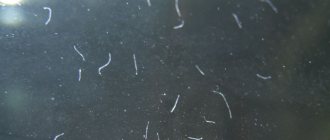
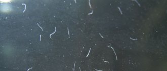
The main food of planarians is protein food. The basis of their diet is small invertebrates, in particular shrimp and crustaceans. They love to feast on the eggs of fish, snails and crustaceans, as well as their food. Often planaria in an aquarium (see photo below) attack adult specimens. They are able to penetrate under their shell and clog their gills, causing suffocation. After which the worms eat the victim.
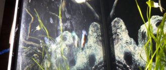
Planaria in a marine aquarium cause no less damage. Due to the colossal speed of reproduction, parasites are able to cover living stones, corals, glass and soil with a continuous crust in a matter of months. Corals covered in planarian secretions begin to suffocate and may eventually die. In addition, in the presence of parasites, the walls of the aquarium take on an unaesthetic appearance.
Reasons for the appearance of Planaria in the aquarium
The appearance of worms can be caused by the following factors:
- Excess feed or untimely removal of remaining food. The food begins to rot, favorable conditions are created;
- The purchased live food turned out to be of poor quality and contaminated;
- At the time of purchase, the vegetation, soil or decor already contained larvae and eggs of parasites;
- The worms were introduced with new fish that were placed in an aquarium without quarantine;
- Too much protein food;
- Cleanliness requirements were not met.
Preventive measures
To prevent the appearance of uninvited guests in the form of planaria, it is necessary to regularly clean the aquarium and prevent it from becoming dirty. The corpses of dead fish and leftover food should not remain in the aquarium for a long time, as the process of rotting will occur.
Due attention should be paid to filters - they need to be washed and cleaned in a timely manner.
It is better to transfer plants and interior elements to the aquarium after pre-treatment with a vinegar solution and thorough rinsing with water.
How to get rid of planaria in an aquarium
It’s not easy to get rid of, but there are methods:
- increase the water temperature - Planaria will begin to crawl out of the ground, you can catch them (this method cannot be called effective, since eggs or larvae may remain in the aquarium);
- reduce feeding of fish, remove remaining food with a siphon (reproduction will stop, it will be easier to get rid of the remaining food);
- some fish, such as Gourami, Melanothenia rainbowfish, Macropods, black-striped Cichlazomas and Bettas, eat Planaria (but reluctantly). The main thing is to put the fish on a hunger strike for several days before adding them to the aquarium;
- traps. To do this, you need to place a vessel with bait in the aquarium. At night, the worms will gather “for a feast” (thanks to their highly developed sense of smell); they can be easily pulled out with a trap. But in this case there is no guarantee that you will get rid of all individuals;
- a quick way is chemical preparations containing fenbendazole. You need to be careful here. Medicines may be safe for shrimp, but harmful to ornamental snails. Another side effect is that dead Planaria begin to rot, and the level of ammonia in the water increases. It is necessary to monitor the parameters more carefully and make changes more often;
- A radical measure is a complete restart of the aquarium.
To get rid of eggs that are difficult to detect, you can use electric current (voltage 12 V).
What you should not do is try to crush or cause any other mechanical injury. So the number of individuals will only increase due to their ability to regenerate.
There are also folk methods of disposal:
- salt (in a ratio of 1g/1l, respectively). It will not be possible to completely destroy it, but the number will be greatly reduced;
- using table vinegar. To do this, you need to make a weak solution (0.2% or 0.5%). After this, you need to rinse all the items in the aquarium with boiled water, change the water or clean it with a filter.
Before using these two methods, it is necessary to remove all animals and vegetation from the container.
Chemicals
How to remove planaria from an aquarium? This question worries many aquarists.
Today, the simplest and most reliable method of getting rid of planaria is to use drugs that contain fenbendazole. These include Flubendazole, Fluvermal, Flubenol or Panacur. The main active ingredient does not harm the permanent inhabitants of the aquarium, but is effective in the fight against planaria.
The recommended dose of fenbendazole is 0.2-0.4 g per 100 liters of water. A day or two after using the drug, all planaria die. Moreover, fenbendazole in the form of a suspension gives a faster effect than its powder counterpart, since the latter is poorly soluble in water. Dead worms are found stuck to the walls of the aquarium. Therefore, the final stage in the fight against parasites will be mechanical cleaning and water replacement.
Interesting Facts
- are able to “learn” at the genetic level by eating their relatives;
- all planarians are hermaphrodites;
- have an extraordinary defensive reaction. If living conditions worsen, the worm falls apart. When the external environment becomes favorable, the Planaria is restored into a whole animal;
- is able to restore the body, even if more than 90% is lost;
- ciliated worms are the only free-living ones, all other flatworms are parasites;
- white Planaria secrete mucus, which is toxic to its inhabitants;
- The only useful function is that flatworms serve as a bioindicator. If the worms have left their shelter in the ground, it is necessary to check the water parameters.
Video about Planaria:
Conclusion
All living things are part of an ecosystem. The appearance of Planaria in an aquarium can not always lead to disaster if you are careful. In small quantities, they do not pose a danger to the inhabitants; they can feed on food particles contained in the water.
To prevent the spread from becoming large-scale, it is necessary to monitor cleanliness: remove leftover food in a timely manner, do regular water changes, clean and rinse the aquarium filter. When purchasing live food, check it carefully, even if it is frozen. Clean plants and decorations periodically.
If the aquatic environment is not clean enough, Planaria will begin to multiply quickly. These eyelash worms are highly resilient, as evidenced by their unique ability to regenerate even from a hundredth part of the body. Therefore, if you allow parasites to multiply uncontrollably, it will be difficult to get rid of them.
Share with friends on social media. networks
10% DISCOUNT FOR BUYERS FROM REGIONS OF RUSSIA AND CIS COUNTRIES FOR AQUARIUMS, FISH, PLANTS, ETC.
Prevention
Since the cause of planaria is the lack of compliance with sanitary and hygienic standards, prevention consists of careful care of the aquarium and monitoring of new inhabitants and new decorations. It is important to frequently change the water in the container, regularly and carefully care for decorations and aeration systems, and also monitor the health of the fish. In this case, the planaria will never disturb the aquarium inhabitants.
An aquarium is a wonderful ecosystem that anyone can recreate in their own home. But, in order for its inhabitants to be healthy, and the appearance of the container itself not to be spoiled by various parasites, due attention must be paid to the care and maintenance of this aquatic world. And then the owner of the aquarium will enjoy the beauty and health of his pets.

