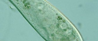
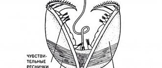
Sessile ciliates of the genus Vorticella, similar to small bells, attached with a long stem to some substrate.
Sometimes they even sit on mollusk shells. The size of such a bell is usually about 50 micrometers, but there are bells up to 170 micrometers.
Under any external influence, the stalk of the ciliate instantly shortens, curling up like a corkscrew, and the bell shrinks into a small ball.
The structure of the stem of the souvoek is quite complicated. Inside it runs a worm-shaped, spirally convoluted cell process, consisting of a bundle of contractile fibers. This bundle is surrounded by mitochondria, which supply energy for the fibers to operate. The contraction of these fibers causes the stem to curl like a corkscrew.
The rest of the stalk is filled with loose extracellular material. This material has elastic properties and straightens the stalk when the contractile fibers relax.
How do Suvoikas eat?
Along the edge of the bell there are three dense rows of cilia: the outer row is horizontal, the two inner ones are vertical. The cilia are in constant motion, and their spirally twisted rows create a flow of water towards the mouth opening. Suvoyki, like slippers, constantly feed, their mouth is always open, and the flow of liquid to it does not stop. Bacteria are the main food for Suvoykas.
Suvoikas spend a significant part of their life in an attached state, and resettlement is carried out using a free-swimming stage - a vagrant. At the bottom of the bell, in front of the stem, a ring of cilia appears; the ciliated rings at the anterior end of the body are retracted inward, and the ciliate is separated from the stalk. Such a wanderer can swim for several hours and then attaches itself to the substrate again. A stalk grows, the rows of cilia at the front end straighten out, and the feeding process begins.
Suctoria sucking ciliates
Rice. Sucking ciliates A - sucking Dendrocometes para¬doxus; 1 - caught prey; 2 - branched tentacles; 3 - contractile vacuole; 4—macronucleus; B — sucking tentacle of Dendrocometes; 1—pellicula; 2— tubules; 3—cytoplasm; B—Sphaerophrya, sucking several ciliates.
Rice.
45. Suvoyki (Vorticella): Sucking ciliates are a small specialized group of predators and parasites, including several dozen species. All suctoria are sessile animals, and therefore in the adult state they are completely devoid of cilia. They are attached in various ways to the substrate, often to the integument of animals, such as crustaceans, using a special leg or sole. Sucking ciliates lack the usual organelles for ciliated ciliates: peristome, mouth, pharynx, digestive vacuoles, etc. Nutrition occurs with the help of special sucking tentacles, which are plasma tubes with a channel leading into the endoplasm (Fig. 46). As soon as any ciliated species or flagellate accidentally touches the sucking tentacle, this protozoan sticks to it and is captured by the sucking ciliate. The latter pulls other tentacles towards the victim. The shell of the protozoan dissolves at the points of attachment of the tentacles, and the cytoplasm of the victim flows through the tubes inward, into the endoplasm of the sucking ciliate. The sucking tentacles can be arranged differently: either radially (Sphaerophrya, Fig. B), or in bunches at the free end of a sitting ciliate, or on special outgrowths - “arms” (Dendrocometes paradoxus; Fig. A, B). Suckers live in both fresh waters and seas. Some of them are predators, eating other ciliates; others parasitize within them. For example, Sphaerophrya sol parasitizes inside the shoe. Asexual reproduction of sucking ciliates occurs by external or internal budding. Buds equipped with a belt of cilia are separated from the mother. They are called tramps. They swim for some time, then attach to the substrate, lose their cilia and turn into typical sucking ciliates. Suckers have a macronucleus and a micronucleus. The sexual process, like in ciliated animals, occurs through conjugation. The presence of cilia and two nuclei in the vagrants, and the presence of the sexual process in adults by sucking by conjugation brings them closer to ciliated ciliates and allows us to believe that sucking ciliates are descended from ciliated ciliates.
Reproduction of sowwok
The formation of vagrants also occurs during asexual reproduction of vagrants. In this case, the ciliate on the stalk divides, and one of the daughter individuals becomes a wanderer and swims away. Under unfavorable conditions, suvoikas are capable of encysting, that is, becoming covered with a dense shell.
The class of Ciliates includes about 6 thousand species. These animals are the most highly organized among the protozoa.
The habitat of ciliates is sea and fresh water, as well as moist soil. A significant number of species of ciliates (about 1 thousand) are parasites of humans and animals.
Let's get acquainted with the morphological and biological features of the structure of ciliates using the example of a typical representative - the slipper ciliate.
Prevention
To avoid infection with a dangerous disease, you should follow basic rules of personal hygiene:
- wash your hands before eating and after using the toilet;
- treat fruits and vegetables with hot water;
- Boil water to make tea or other drinks.
On a public scale, measures should be taken:
- to combat contamination of human habitations with pig excrement;
- to improve occupational hygiene and to prevent possible harmful effects on the health of people working with animals on farms;
- for timely diagnosis of the disease and treatment of people infected with balantidiasis.
Section: Protozoa Please rate how much you liked this article:
Pressing the social button gets rid of all kinds of parasites, cleanses your body, makes you healthy, beautiful, cheerful and full of vitality.
External and internal structure of the ciliate slipper
The slipper ciliate has a size of about 0.1-0.3 mm. The body shape resembles a shoe, which is why it got its name.
This animal has a constant body shape, since the ectoplasm is compacted on the outside and forms a pellicle . The body of ciliates is covered with cilia. There are about 10-15 thousand of them.
A characteristic feature of the structure of ciliates is the presence of two nuclei: large (macronucleus) and small (micronucleus). The small nucleus is associated with the transmission of hereditary information, and the large nucleus is associated with the regulation of vital functions. The slipper ciliate moves with the help of cilia, with the anterior (blunt) end forward and at the same time rotates to the right along the axis of its body. The high speed of movement of the ciliate depends on the paddle-like movement of the cilia.
Diagnosis and treatment
Diagnosis of the presence of the ciliate Balantidium coli in the body is carried out using a native (preserving the natural structure and color of the test material) smear or scraping from the affected area of the intestine, taken using the sigmoidoscopy procedure. The ciliates themselves are detected quite easily due to their large size, characteristic shape and high mobility. Cysts are more difficult to detect. They can be recognized using solutions stained with Lugol's preparation. Typically, a small amount of balantidia is observed in smears, and to make an accurate diagnosis it is necessary to undergo analysis several times. Important information when making a diagnosis is the patient’s place of residence, the proximity of farms and places where livestock are kept.
Diagnosis of dangerous bacteria is carried out in several ways:
Research under a microscope
In this case, a native smear is examined. Balantidia are clearly visible under magnification, since their length is about 75 µm and thickness is about 40 µm. To detect it, a small magnification of the microscope is sufficient. To study the microorganism in more detail, it is worth removing excess liquid from the preparation. The causative agent of the disease will slow down and it can be examined in more detail. At this magnification, cilia are visible that evenly cover the egg-shaped body of the ciliate. In the center, a bean-shaped body is visible - the vegetative nucleus (macronucleus). It is surrounded by a turbid granular liquid - endoplasm. The next layer is ectoplasm and cytoplasm, which limits the cell from the external environment. Vacuoles are located in the front and back of the body. They look like light balls that appear and disappear.
Heidenhain method
When studying a drug using the Heidenhain method, the signs of microorganism manifestation are the same as when examined under a microscope. Observation is also carried out at a relatively low magnification. Balantidia are easily detected due to the internal structure of the cell. A slight difference is observed in the absence of cilia in the medium in question.
Culture method
To study balantidia using this method, Rice medium is used.
Treatment of a patient with balantidiasis should be carried out under the strict supervision of a doctor or in a hospital. To improve well-being and speed up recovery, the patient is prescribed the following medications:
Specific dosages are prescribed by a specialist. With modern therapy, the prognosis for the outcome of the disease is favorable. Without recourse to antiparasitic therapy, the mortality rate from the disease reaches only 12%.
Nutrition and excretory organs
The nutritional organelles of the slipper ciliate are: the preoral cavity, the cellular mouth and the cellular pharynx. Bacteria and other particles suspended in water, along with water, are driven by perioral cilia through the mouth into the pharynx and enter the digestive vacuole.
Nutritional organs of ciliates-slippers
Having filled with food, the vacuole breaks away from the pharynx and is carried away by the current of the cytoplasm. As the vacuole moves, the food in it is digested by digestive enzymes and absorbed into the endoplasm. Then the digestive vacuole approaches the powder and undigested food remains are thrown out. Ciliates stop feeding only during the breeding season.
The organelles of osmoregulation and excretion in the slipper are two contractile, or pulsating, vacuoles with actuator canaliculi.
Thus, ciliates, in comparison with other protozoa, have a more complex structure:
- Constant body shape;
- the presence of a cellular mouth;
- presence of a cellular pharynx;
- powder;
- complex nuclear apparatus.
Reproduction of ciliates. Conjugation process
Ciliates reproduce by transverse division, in which nuclear division first occurs. The macronucleus divides amitotically, and the micronucleus divides mitotically.
From time to time they undergo the sexual process, or conjugation . During this, two ciliates come closer and closely attach to each other with their mouth openings. At room temperature in this form they float for about 12 hours. Large nuclei are destroyed and dissolved in the cytoplasm.
Reproduction of ciliates
As a result of meiotic division, migrating and stationary nuclei are formed from small nuclei. Each of these nuclei contains a haploid set of chromosomes. The migrating nucleus actively moves through the cytoplasmic bridge from one individual to another and merges with its stationary nucleus, that is, the process of fertilization occurs. At this stage, each shoe forms one complex nucleus, or synkaryon, containing a diploid set of chromosomes. Then the ciliates disperse, their normal nuclear apparatus is restored again, and they subsequently multiply intensively by fission.
The process of conjugation contributes to the fact that the hereditary principles of different individuals are combined in one organism. This leads to increased hereditary variability and greater resilience of organisms. In addition, the development of a new nucleus and the destruction of the old one is of great importance in the life of ciliates. This is due to the fact that the basic life processes and protein synthesis in the body of ciliates are controlled by a large nucleus.
general characteristics
The type of ciliates unites a large number of species (over 6000) of the most highly organized protozoa. They are characterized by the presence of cilia, which are usually present in large numbers. Cilia serve as organelles of movement; they can stick together to form more complex organelles. Some sucking ciliates have cilia only in the early stages of the life cycle. All ciliates are characterized by nuclear dualism, i.e. duality. This means that they have at least two nuclei that differ in both size and function. One of the nuclei, much larger, is called a macronucleus, and the second, small one, is called a micronucleus. Some types of ciliates have several micro- and macronuclei. The micronucleus serves as the sexual, or generative, nucleus, playing a major role in the sexual process. Macronucleus is a somatic, or vegetative, nucleus that regulates all life processes except the sexual process. Asexual reproduction of ciliates occurs by transverse division. The sexual process in ciliates occurs in a unique way, in the form of conjugation, which is not observed in protozoa of other classes. Conjugation consists of the temporary bringing together of two individuals and the mutual exchange of parts of their micronuclei. Ciliates are inhabitants of mainly fresh water bodies, but are also found in brackish water and in the seas; some species have adapted to existence in moist soil. Among the ciliates there are many parasites (about 1000 species) of invertebrate and vertebrate animals. The class is divided into two classes:
- Ciliated ciliates (Ciliata)
- Sucking ciliates (Suctoria).
The importance of ciliates in nature and human life
It has been established that ciliates play a significant role in the cycle of substances in nature. Various species of larger animals (fish fry) feed on ciliates.
They serve as regulators of the number of unicellular algae and bacteria, thereby cleaning water bodies.
Ciliates can serve as indicators of the degree of contamination of surface waters—water supply sources.
Ciliates living in the soil improve its fertility.
Man breeds ciliates in aquariums to feed fish and their fry.
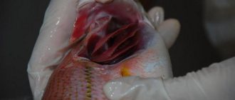
In a number of countries, human and animal diseases caused by ciliates are widespread. Particularly dangerous is the ciliate balantidium, which lives in the intestines of pigs and is transmitted to humans from the animal.
| Kingdom | Animals |
| Sub-kingdom | Unicellular |
| Type | Ciliates |
Routes of human infection
Balantidium coli is a dangerous species of ciliate that lives on the inner walls of the intestines of humans and pigs. Infection can occur due to failure to comply with personal hygiene rules. Balantidia cysts enter the rectum and may not manifest themselves for a long time. Under the influence of various factors, they are activated and destroy the intestinal mucosa, forming ulcers. In the intestines of pigs, balantidia easily cyst, while the human intestines can bear the parasite for a long time and not show its activity in any way.
The most common places of infection are farms and private farms where pigs or livestock are raised. Farm workers themselves are at greater risk than the rest of the population. The fact is that cysts are quite tenacious and can remain in an animal’s droppings for a long time. Cysts can persist in pig excrement for up to several weeks. In vegetative form, at room temperature, they die after 2-3 days. The cysts themselves can be carried by birds and insects, ending up on vegetables and fruits growing dangerously close to livestock. They can also be transmitted through water or through tactile contact with a contaminated object or an already sick person. When a person is infected, characteristic symptoms appear.
Habitat, structure and movement
The slipper ciliate lives in shallow, stagnant bodies of water. This unicellular animal, 0.5 mm long, has a spindle-shaped body, vaguely reminiscent of a shoe. Ciliates are constantly in motion, swimming with their blunt end forward. The movement speed of this animal reaches 2.5 mm per second. On the surface of the body they have organelles of movement - cilia. There are two nuclei in the cell: the large nucleus is responsible for nutrition, respiration, movement, metabolism; The small nucleus is involved in the sexual process.
The structure of the ciliate slipper
The body of a ciliate is more complex. The thin elastic shell covering the outside of the ciliate maintains the constant shape of its body. This is also facilitated by well-developed supporting fibers, which are located in the cytoplasm layer adjacent to the membrane. There are about 15,000 oscillating cilia on the surface of the ciliate's body. At the base of each cilium lies a basal body. The movement of each eyelash consists of a sharp stroke in one direction and a slower, smooth return to its original position. The cilia oscillate approximately 30 times per second and, like oars, push the ciliates forward. The wave-like movement of the cilia is coordinated. When a slipper ciliate swims, it slowly rotates around the longitudinal axis of the body.
Symptoms and complications
The scientific name for the parasite is balantidiasis. When suffering from balantidiasis, the colon is affected by erosions and ulcers. As a result, swelling, suppuration of the lesion and tissue necrosis occurs. In advanced cases, the disease can be fatal.
Pathological processes begin as a result of the proliferation of microorganisms in the walls of the colon; infection of the final part of the small intestine and the formation of ulcers are possible. The disease can occur in acute or chronic form. Patients with a chronic form of the disease are more often observed.
In any form, a characteristic sign will be the appearance of bloody diarrhea with mucus and a foul odor, or the occurrence of colitis, accompanied by the release of semi-liquid mucous diarrhea without blood. The chronic form of the course may not manifest itself in the form of dysentery at all, but may be in remission. In this case, patients believe that they have a mild illness and do not turn to specialists. With a long-term onset of the disease, periods of remission may be shortened, and acute conditions will manifest themselves more strongly. At such moments, the chance of death is much higher.
The development of the disease is usually divided into three stages:
- incubation period;
- acute period;
- chronic balantidiasis.
A patient with balantidiasis experiences the following symptoms: loss of appetite, headache, fever, moderate fever (or heat), weakness. Along with the main signs, characteristic symptoms of the disease may also appear: flatulence, abdominal pain, diarrhea; if the rectum is affected, tenesmus (false urge to defecate, accompanied by painful sensations) may be observed. The patient's stool contains blood and mucus. There may be a dry tongue, painful feelings in the liver area (according to tactile sensations, it increases). In severe cases of the disease, severe fever begins, frequent nausea, and foul-smelling diarrhea with blood and mucus appear. Such patients lose weight very quickly, and a week later they develop cachexia (depletion of the body).
Life processes
Nutrition
The slipper and some other free-living ciliates feed on bacteria and algae.
Reaction of ciliates to food
The thin elastic membrane (cell membrane) covering the ciliate from the outside maintains a constant body shape. There are about 15 thousand cilia on the surface of the body. There is a depression on the body - a cellular mouth, which passes into a cellular pharynx. At the bottom of the pharynx, food enters the digestive vacuole. In the digestive vacuole, food is digested within an hour, first with an acidic and then with an alkaline reaction. Digestive vacuoles move in the body of the ciliate by a current of cytoplasm. Undigested remains are thrown out at the posterior end of the body through a special structure - a powder located behind the mouth opening.
Reproduction
Asexual
Ciliates usually reproduce asexually - by dividing in two. The nuclei are divided into two parts, and each new ciliate contains one large and one small kernel. Each of the two daughters receives part of the organelles, while the others are formed anew.
Sexual
With a lack of food or a change in temperature, ciliates proceed to sexual reproduction, and then can turn into a cyst.
During the sexual process, the number of individuals does not increase. Two ciliates are temporarily connected to each other. At the point of contact, the shell dissolves and a connecting bridge is formed between the animals. The large core of each ciliate disappears. The small nucleus divides twice. Each ciliate produces four daughter nuclei. Three of them are destroyed, and the fourth is divided again. As a result, two cores remain in each. An exchange of nuclei occurs along the cytoplasmic bridge, and there it merges with the remaining nucleus. The newly formed nuclei form a large and small nuclei, and the ciliates disperse. This sexual process is called conjugation. It lasts about 12 hours. The sexual process leads to renewal, exchange between individuals and redistribution of hereditary (genetic) material, which increases the vitality of organisms.
Class Ciliated ciliates (Ciliata)
Slipper ciliates, trumpeter, suvoiki, balantidium
The slipper ciliate (Paramecium caudatum) (Fig. 1A) lives in fresh water bodies. The cell membrane contains a flexible, thin pellicle that gives the body a permanent shape. Under the pellicle there is a transparent layer of ectoplasm, which contains supporting fibrils, basal bodies of cilia and trichocysts. Trichocysts are small round organelles that, under the influence of an irritant, emit thin pointed filaments. Trichocysts are organelles of defense and attack. The entire body is covered with cilia. On the lateral surface of the body there is a depression - the peristome (perioral funnel). At the bottom of the peristome is a cytostome (cell mouth), which passes into the cytopharynx (cell pharynx). The cytopharynx is immersed in the endoplasm. The slipper ciliate feeds on bacteria, algae, and suspended particles of organic matter. The ciliary apparatus of the perioral funnel directs the food particle through the cytostome into the cytopharynx.
In the endoplasm, this food particle is packaged into a membrane, under which numerous lysosomes “pour out” their enzymes: a digestive vacuole is formed. The digestive vacuole does not remain in place, but, entering the endoplasmic currents, completes a rather complex path called cyclosis of the digestive vacuole. At the beginning of cyclosis, the environment in the vacuoles is acidic, and at the end it is alkaline. Undigested residues are thrown out through the cytoproct (anal pore, powder). The release mechanism is homologous to exocytosis.
The slipper ciliate has two contractile vacuoles, each of which consists of a reservoir and 5–7 afferent canals. First, the adductor channels are filled with liquid, from which the contents are pushed into the reservoir, then from the reservoir through the excretory pore to the outside.
The endoplasm contains one micronucleus and one macronucleus. The macronucleus is large, bean-shaped, regulates the vital activity of the cell, and is polyploid. Micronucleus - small diploid - controls the reproduction process. Unlike macronuclei, RNA synthesis does not occur in micronuclei.
rice. 1. Ciliated ciliates. A - ciliate-slipper, B - trumpeter: 1 - peristome, 2 - cytostome, 3 - cytopharynx, 4 - digestive vacuole, 5 - macronucleus, 6 - micronucleus, 7 - reservoir of the contractile vacuole, 8 - adductor channels of the contractile vacuole, 9 - cilia, 10 - trichocysts, 11 - cytoproct.
When unfavorable conditions occur, the slipper ciliates transform into cysts.
Reproduction is asexual (transverse cell division in two), which alternates with the sexual process. Recently, a number of authors have called this sexual process, found only in ciliates, a primitive form of sexual reproduction such as conjugation. During asexual reproduction, the micronucleus divides by mitosis, the macronucleus by amitosis. Conjugation of ciliates and slippers is a complex process that lasts several hours. The following stages can be distinguished in conjugation (Fig. 2).
rice. 2. Conjugation in ciliates - slippers.
- The connection of two ciliates (conjugants) with each other by peristomal areas with the formation at the point of contact of a cytoplasmic bridge connecting both ciliates.
- Dismantling of macronuclei, division of micronuclei by meiosis to form four haploid nuclei.
- Destruction of three haploid nuclei, division of the fourth by mitosis. Of the two haploid nuclei formed, one will remain in place, the second will move along the cytoplasmic bridge to another ciliate. The first core is conventionally called female, the second - male.
- Exchange of male nuclei, the consequence of which is the recombination of genetic information.
- Fusion of male and female nuclei to form a diploid syncarion, divergence of ciliates. After divergence, ciliates are called exconjugants.
- The synkaryon divides three times by mitosis, the formation of eight diploid nuclei, of which four are micronuclei, four are future macronuclei.
- Dismantling of three micronuclei, formation of an infusoria with five nuclei: one is a micronucleus (diploid), four are macronuclei, also diploid.
- The division of each of the exconjugants in two, with the generative nuclei dividing by mitosis, and the macronuclei separating in pairs into daughter cells. Formation of four daughter ciliates, each of which has one micronucleus and two macronuclei.
- The division of each of the four daughter ciliates in two, again the micronuclei divide by mitosis, and the macronuclei disperse into daughter ciliates. As a result, four daughter ciliates are formed from one mother ciliate, each of which has one macronucleus and one micronucleus. The micronucleus remains diploid. The macronucleus becomes polyploid because DNA is replicated before each divergence.
rice. 3. Autogamy in ciliates - slippers.
With prolonged asexual reproduction, as a result of amitotic division of the macronucleus (random distribution of DNA), the vital functions of the ciliate may be disrupted. To restore the normal set of chromosomes and, therefore, normal life activity, a process called autogamy occurs (Fig. 3). Autogamy can be divided into the following stages.
- Dismantling of the macronucleus, division of the micronucleus by mitosis three times to form eight haploid nuclei.
- Dismantling of six haploid nuclei. Fusion of the two remaining nuclei to form a diploid synkaryon.
- Synkaryon division, two nuclei become micronuclei, two become macronuclei.
- Division of ciliates to form two individuals, each of which has a normal micronucleus and macronucleus.
The Trumpeter (Stentor polymorphus) lives in fresh water bodies. It has a characteristic funnel-shaped body (Fig. 1B). It can swim freely and can attach itself to the substrate with the narrow posterior end of the body, releasing short pseudopodia. At the anterior end of the body, expanded in the form of a bell, there is a peristomal field. The body of the trumpeter is covered with small cilia. Along the outer edge of the peristomal field are membranellae formed by the fusion of a number of cilia. The trumpeter has one contractile vacuole, consisting of a reservoir and two afferent canals.
In the endoplasm along the body there is a long, clearly visible macronucleus, next to the macronucleus there are several micronuclei.
Suvoiki (Vorticella sp.) are inhabitants of fresh water bodies. Stay in groups. A long thin stalk extends from the rear end of the body, with which the suvoika is attached to plants, mollusk shells, etc. The stalk is capable of sharply twisting into a spiral. The cilia of Suwoikas are located only around the oral funnel in two rows, forming a left-handed double helix. Suvoyki have one contractile vacuole without adductor canals, one horseshoe-shaped macronucleus, one micronucleus.
rice. 4. Suvoyki: 1 - beginning of division, 2 - separation of the "vagrant", 3 - "vagrant", 4 - conjugation.
rice. 5. Balantidium coli
Reproduction is asexual, alternating with conjugation. As a result of asexual reproduction, free-swimming “vagrants” are formed (Fig. 4). “Tramps” ensure the resettlement of the sows. After swimming for some time, the “vagrants” descend to the substrate and form a stalk. Conjugation is anisogamous: one of the conjugant is a suvoyka with a stem, the second conjugant is a “vagrant”.
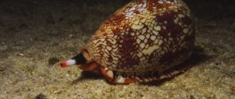
Balantidium coli (Fig. 5) parasitizes the large intestine of humans and pigs. In humans it causes a serious disease - balantidiasis. The invasive stage is cysts, which can be found on unwashed vegetables, fruits, dirty hands, or in unboiled water. Balantidium can live in the intestines without causing pathological changes (cyst carriage). In some cases, balantidia are embedded in the intestinal wall, and deep bleeding ulcers form at the sites of penetration. Balantidiasis is characterized by bloody diarrhea and pain in the intestines. The main sources of invasion are pigs infected with balantidia.
►