Treatment of lerneosis in fish
Lerneosis to treat . Aquarists use various drugs and various methods to treat this disease. You can use the following method for treating lerneosis .
For sick fish, prepare a quarantine aquarium with optimal conditions and add a solution of table salt to it at the rate of approximately 1 tablespoon of salt per 10 liters of water. The fish are kept in a saline solution until they are completely recovered and the wounds are healed after the procedure for removing crustaceans from the body of sick individuals (2-3 weeks).
It is a good idea to use hydrochloride instead of salt in a quarantine aquarium at the rate of 2 mg of the drug per 10 liters of water. The duration of treatment in the hydrochloride solution should be four days.
Before placing fish affected by lerna in a quarantine aquarium, they are caught from the general aquarium and, carefully clamped in a wet cotton swab, the crustaceans are very carefully removed from the body with thin tweezers with smooth edges: if part of the crustacean remains in the body of the fish, the wound will rot.
Fish from the quarantine aquarium are periodically caught and lotions are made with a swab dipped in a solution of potassium permanganate of a not too saturated dark pink color, or the fish are bathed in manganese baths (of medium color).
In order to prevent the recurrence of lerneosis and destroy all lerne eggs, when there are no fish in the general aquarium, it is disinfected, for example, with trypaflavin. This drug is sold in pet stores and is used according to the scheme given in the instructions for use.
Ichthyophthiriasis
This is one of the most dangerous ectoparasitic diseases that can cause massive waste of fish, especially juveniles. Almost all types of fish get sick. The disease is caused by ciliary infusuria, the name of which is translated from Latin as “Fish louse of many children.” The parasite develops and matures under the skin of the fish, and therefore it is resistant to many drugs that are effective for other diseases. Having reached maturity, the parasitic ciliate leaves the fish, sticks to underwater objects, and forms a cyst. After repeated division, several thousand daughter cells are formed in it. These cells then go out into the water and float freely for 2-3 days. If they manage to attach to the fish, they are embedded under the skin, where they develop.
Sick fish are weakened, stay in the upper layers of water, and react poorly to external stimuli. A small white rash similar to semolina is noticeable on the surface of the body and gills. The diagnosis is made only after a microscopic examination of scrapings from the surface of the skin and gills, since a “white rash” can appear with some myxosporidiosis, and can also be a manifestation of the “nuptial plumage” of male carp during the spawning period.
The fight is difficult because the parasite is located under the skin of the fish. Treatment is carried out only under the guidance of an ichthyopathologist. Prevention consists of preventing the entry of trash fish into reservoirs, pools, and cages where industrial cultivation is carried out. Transportation and transplantation of fish must be carried out using such preparations as malachite green, violet “K”, basic bright green, potassium permanganate.
Pathogen
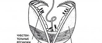
The body of a sexually mature female is elongated - up to 10-16 mm, cylindrical in shape, undivided, somewhat expanded towards the posterior end. At the head end there are four outgrowths (a pair of branched and a pair of unbranched), with the help of which the lernaea are embedded in the body of the fish. There are five pairs of bibranched swimming legs. The egg sacs are paired, elongated, each containing from 300 to 700 eggs.
Trichodinosis, apiozomosis, costiosis, scyfidiasis, trichophriasis
The diseases are caused by parasitic ciliates that develop on the surface of the body and gills of almost all cultivated fish species. When there are 1-5 parasites in the microscope field of view (10x10), clinical signs of the disease usually do not appear. With a population of 10-15 specimens/p.z. cause anxiety in fish, increased sliming of the integument, and a bluish or gray coating appears on the body of the fish. The diagnosis is made after microscopy of scrapings. Treatment is selected taking into account environmental conditions and the condition of the fish. There are several effective drugs, which include malachite green, brilliant green, violet “K”, potassium permanganate, table salt, formaldehyde solution, etc.
Development
In the egg sacs of a sexually mature female, young copepods (nauplii) with three pairs of legs develop in the summer and go out into the water. In water they go through three nauplial and five copepodite stages, molting each time. At the fifth stage of development, differentiation of the sexes occurs, females and males are formed. Soon after copulation, the males die, and the females attach to the skin of the fish, penetrate the tissues and reach sexual maturity. Lernae are very prolific, and during the summer period there is a repeated change of new generations of crustaceans. The speed of their development depends on the temperature. The autumn generation of crustaceans overwinters on fish. Reproduction of lernae is observed only in fresh water.
Philometroidosis of carp
Philometroidosis is caused by roundworms called nematodes. The main host of the parasite is carp, the intermediate host is cyclops. Female phylometras reach a length of 90 - 160 mm. They are localized in the carp's scale pockets in the area of the head, pectoral fins, and behind the gill covers. Males are much smaller than females and are usually found in the walls of the swim bladder.
When cleaning fish from scales, female phylometers look like pink-red rings with weak mobility. Although phylometra are not dangerous to humans, their detection in fish causes a negative reaction in the person preparing the fish. The buyer usually returns such fish to the sellers. The main cause of the disease is the transportation of fish from unfavorable fish farms to prosperous ones. Treatment of fish is possible using a number of drugs that have a high therapeutic effect.
Epizootological data.
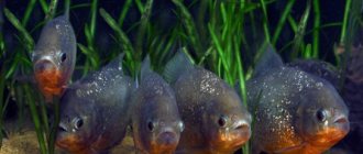
Lerneosis is widespread both in pond farms and in lake-type reservoirs. The disease manifests itself in the summer, more often in old silted ponds, when they are kept unsanitary. The most susceptible to the disease are fry and underyearlings of crucian carp, carp, carp, buffalo and black carp. Infected fish begin to be detected at the end of April in reservoirs in the southern zones and in mid-summer in the central zone. The appearance of clinical signs and death are observed at the end of summer. The parasite develops most intensively at temperatures above 23°C.
Inflammation of the carp swim bladder (SVB)
The cause of the disease is not fully understood. The most substantiated point of view is the viral nature of the disease. The infection is transmitted mainly through direct contact between sick and healthy fish. Apparently, the pathogen can also be transmitted through water and soil.
Carp, carp and their hybrids are affected. Occasionally, cases of the disease have been reported in silver carp, pike, grass carp, crucian carp and pike. The main symptom of this disease is damage to the walls of the swim bladder. Inflammation often affects other internal organs. Fish of different age groups are susceptible to the disease. Fingerlings of carp suffering from STD usually die during wintering. In the acute form of the disease, massive waste is possible. Over time, fish develop immunity to this disease, and it gradually subsides. However, this is only possible if quarantine restrictions are observed. There are no specific drugs against this disease.
Symptoms
Lernae penetrate the skin and reach muscle tissue, penetrating deeply into it, and are located over the entire surface of the body. In places where the crustacean invades the tissue, inflammation, swelling, and hyperemia develop, followed by the formation of ulcers with a white narrow rim. Pathogenic bacteria and fungi inhabit the affected areas. Due to the impregnation of tissues with bloody exudate, the scales rise, become deformed and fall out. Sick fish refuse food, are exhausted, move slowly, accumulate on the water flow and die, especially young of the year carp and buffalo. The prevalence of crustaceans is high; there are dozens of parasites on the skin of fish. Fish affected by lernaea become spreaders of the disease the following year. The source of invasion is also the larval stages of lerna, penetrating into ponds with water from unfavorable head or other water supply sources. In pond farms in Japan, lernaea settles in the oral cavity of eels, causing ulceration, curvature of the jaws and significant death.
Saprolegniosis
Saprolegniosis is one of the most common fish diseases. It is believed that saprolegniosis is a secondary disease that occurs at the site of traumatic injuries on the fish’s body. In addition to trauma, saprolegniosis appears as a concomitant disease with other diseases, both infectious and invasive. The causative agent of the disease is lower fungi, mainly from the genus Saprolegnia, which are very widespread in nature. Saprolegniosis affects almost all freshwater fish that have been exposed to one or another effect or found themselves in unfavorable living conditions. Saprolegniosis often occurs in carp fish farms as a result of careless handling of fish, when kept in concrete cages, as a result of trauma during fishing, loading and unloading of live fish. The hyphae of the fungus penetrate the damaged tissues of the muscles, gills, and skin of fish, destroying the tissue. On the surface of the body, the fungus forms a coating similar to dirty cotton wool. Prevention is the main way to prevent saprolegniosis. All technological operations must prevent injury to fish. For preventive and therapeutic purposes, you can use drugs such as malachite green, brilliant green, and table salt. A type of saprolegniosis is Staff's disease. It manifests itself mainly in carp underyearlings during wintering. In this disease, fungi develop in the nasal cavities of fish. Fungal mycelium in the form of a cotton wool-like mass covers the head of fish, and fungal hyphae can grow into brain tissue. Staff's disease usually occurs in winter at very low water temperatures. Although there is evidence that this disease also occurs at temperatures of 5-6 degrees. Saprolegniosis of eggs is the scourge of hatcheries. Saprolegniosis affects the eggs of many fish species incubated in artificial conditions, mainly salmon and whitefish. The disease primarily affects dead eggs and quickly spreads to healthy eggs. Selection of dead eggs is a labor-intensive but effective method of preventing saprolegniosis. To combat this disease, malachite green, methylene blue, and brilliant green are widely used. Treatment of water with ultraviolet lamps and ozonation of water also prevent the development of saprolegniosis.
Pathogenesis and pathological changes
The pathogenic effect is reduced to tissue dysfunction, inflammatory processes in muscles, internal organs, especially in the liver. The secretion of the poisonous gland of crustaceans has a negative effect on the general condition of the body, the composition of the blood changes. Disintegration of the affected tissues is observed, focal traumatic hepatitis develops with damage to the liver tissue by crustaceans.
DISEASES OF ZANIO PINK: DESCRIPTION, PREVENTION, TREATMENT, SYMPTOMS, PHOTOS
CICHLID DISEASES: DESCRIPTION, PHOTOS, TREATMENT, TYPES
HOW TO TREAT FISH WITH SALT IN THE AQUARIUM?
BARBUS DISEASES: SYMPTOMS, TREATMENT, PHOTOS
NUTRITIONAL DISEASES
| In industrial aquaculture with the transition to highly intensive forms of fish farming (in cages, pools, etc.), nutritional diseases cause damage. They are divided into 2 groups. The first includes diseases associated with the use of unbalanced feed in terms of fat, protein, carbohydrate, mineral and vitamin composition. The second group includes diseases that occur in fish as a result of consuming low-quality feed, contaminated with microorganisms (bacteria or fungi), their metabolic products, or containing oxidized fats. Nutritional diseases occur in fish of different species and ages. They reduce the growth rate of fish and can cause their death. The peculiarities of their manifestation require not only general, but also special approaches to their prevention. |
FISH DISEASES AND THEIR DANGERS TO HUMAN HEALTH
IS SICK FISH DANGEROUS? Often, fishermen in a reservoir pay attention to the unusual behavior of fish and changes in their appearance. This is evidence that the fish is sick. What contributes to the spread of fish diseases? Most often, it is the release of juveniles into a reservoir that have not undergone a veterinary ichthyopathological examination, as well as the use of live bait caught in “unfavorable” reservoirs. Every fisherman should know the main signs of the most common diseases in order to protect himself from disease and water bodies from infection. Most diseases transmitted to humans from fish are caused by helminths that live in the intestines of fish. Fish diseases: black dots What do small black dots mean on the body of caught fish - roach, bream and other species?
Is it possible to eat such fish? INKSPOT DISEASE (POSTODYPLOSTOMISIS)
In recent years, the distribution areas of many fish diseases have expanded.
This is due both to the deterioration of the ecological situation, which leads to a decrease in the overall resistance of fish to diseases, and to changes in the boundaries of the ranges of various animal carriers of diseases. Black dots on the body of fish, as far as can be judged from the available descriptions of concerned fishermen, are a sign of just one of these diseases. This is post-diplostomosis.
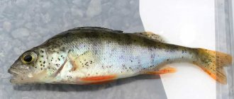
This disease occurs in carp, silver carp, roach, roach, perch and other fish. Affected fish have noticeable black dots scattered throughout the body, on the fins, gills, and cornea of the eyes. Each point is a place in which a capsule with a helminth larva is located. The development cycle of the causative agent of post-diplostomosis is similar to the development cycle of diplostoma. The main host of the parasite is the heron, the first intermediate host is the mollusk, and the second is fish.
More details:
It is caused by the larvae of the parasitic worm Postodiplostomum cuticola from the class of digenetic flukes. An adult worm has a flat body 1.5 mm long and 0.5-0.9 mm wide, but it itself is not directly related to the appearance of blackheads. It parasitizes the intestines of fish-eating birds such as tree frogs or herons. In the intestines of birds, worms produce eggs from which larvae emerge. The larvae enter the water and penetrate the body of the intermediate hosts - mollusks. In the mollusk, asexual reproduction of the larvae occurs, as a result of which larvae are obtained again, but of a different structure - cercariae. So they are parasites on fish.
Where do blackheads come from?
Cercariae enter the water and penetrate the skin and muscles of fish. At the same time, in the subcutaneous tissue of the fish at a depth of 1.5-2 mm around the larva, a connective tissue capsule is formed, in which the black pigment hemomelanin is deposited - a product of the breakdown of blood cells and pigment cells of the skin of the fish. This is how “black spots” appear.
Postodiplostomosis occurs everywhere.
The appearance of this disease may be associated with the spread of mollusks that carry the disease or with the negative impact of pollution of water bodies.
As can be seen from the description of the life cycle of postodiplostomum, humans do not participate in it in any way - they are neither an intermediate nor the final host of the parasite. While in the body of the fish, cercariae do not emit toxins dangerous to humans. According to the relevant GOSTs, if commercial fish has single black spots on its body, it is allowed for sale without any special processing. So fish affected by post-diplostomosis can be eaten. With one caveat.
Fish must be properly prepared (cooked, fried, salted) to exclude the possibility of any other parasitic organisms remaining in it in a viable state. After all, many parasites that can infect humans do not manifest themselves either as black spots or any other noticeable signs
Can a person get infected from fish? Yes maybe. You should KNOW that poorly cooked or undercooked fish not only causes poisoning, but can also be a source of helminth infection for humans and domestic animals. Barely visible to the eye, worm larvae found in apparently healthy fish, once ingested by humans and fish-eating animals, cause serious diseases - Opisthorchiasis and Diphyllobothriasis. Women who process the fish themselves and taste it raw are more often infected.
THINGS TO REMEMBER
Diagnosing a particular fish disease is quite difficult, and ichthyopathologists must do this.
But in some cases, amateur fishermen may encounter diseases that have a characteristic clinical manifestation, and pathogens that are visible even to the naked eye. Such diseases include ligulosis, ink-spot disease (postodiplostomosis), diplostomosis, phylometroidosis, etc.
OPISTHROCHISIS
The disease is caused by small trematodes
- opisthorchis parasites in the bile ducts of the liver, in the pancreas, in the gall bladder of humans and carnivores.
Sexually mature opisthorchises are 6-14 mm long and 1.2-2 mm wide.
In the human or animal body (the main host), opisthorchises secrete eggs that enter the intestines with bile, and then with feces into the external environment.
Opisthorchis eggs are swallowed by a freshwater mollusk (the first intermediate host), in whose body the parasite undergoes further development, and ultimately, from one egg swallowed by the mollusk, many cercariae appear - the larval stages of the parasite. Cercariae emerge from mollusks into the water and actively invade the body of fish (the second intermediate host). In fish, cercariae are localized in the muscles, become covered with a membrane, and turn into metacercariae, which can already cause infection of the main host if they enter its gastrointestinal tract alive. This occurs when eating raw, poorly processed fish. Opisthorchis larvae
infect roach, ide, carp, bream, dace, tench, silver bream, rudd, gudgeon, asp, podust, and minnow.
They do not occur in salmon, sturgeon, or marine fish. Opisthorchises can live in the body of the main host for a long time, and they cause significant harm to it. With opisthorchiasis -
headache, aching pain in the stomach, short-term fever.
The pain in the pit of the stomach increases every day and does not depend on food intake. Eventually the person becomes unable to work. Treatment of opisthorchiasis is associated with a number of difficulties and should only be carried out under the guidance of a doctor. Opisthorchiasis is a natural focal disease that is common where the intermediate host, the mollusk bithinia, lives.
This disease has been recorded in Western Siberia, Kazakhstan, the Perm region, the Volga region, in the basins of the Ob, Irtysh, Volga, Don, Neman, Kama, Dnieper rivers, etc. The carriers of opisthorchis larvae are ide, dace, roach and a number of other fish.
Prevention measures The main cause of opisthorchiasis infection is eating raw, contaminated fish. Conventional cooking -
boiling or frying for 25-30 minutes - can completely eliminate the threat of opisthorchiasis infection.
When salting,
the salt concentration should be at least 14% of the weight of the fish; salting should be carried out for 2 weeks.
Severe freezing
can also destroy opisthorchiasis pathogens.
The temperature must be maintained at -18...-20 °C during the day. In many foci of opisthorchiasis, infection of the population is observed. There are frequent cases of infection with opisthorchiasis and a particularly severe course of the disease among visitors who find themselves in the center of opisthorchiasis. They do not have the partial immunity that local residents already suffering from opisthorchiasis have. People traveling to areas with opisthorchiasis need to remember this.
Under no circumstances should raw fish or its entrails be fed to dogs, cats or other carnivorous animals. They not only themselves become ill with opisthorchiasis, but also become a source that supports the disease in the area and contribute to its wider spread.
DYPHYLLOBOTRIOSIS Diphyllobothriasis is a helminthic disease of humans and carnivores. The causative agents of this disease are
flat parasitic worms
(cestodes).
Like opisthorchises, the development cycle of these helminths is complex, involving two intermediate hosts.
Mature helminths parasitize the human intestine, and they can reach extremely long lengths (up to 10 m).
A sexually mature helminth releases eggs, which are released into the external environment with feces.
If the eggs end up in water, they develop, and after a few days the larvae emerge from the eggs. Cyclops and diaptomus (small crustaceans that fish feed on) swallow the larvae, and after 3-4 weeks they turn into procercoids. If Cyclops or Diaptomus are eaten by fish, the development of the parasite continues.
From its intestines, procercoids enter the liver, muscles, gonads and other internal organs and tissues of the fish and turn into plerocercoids.
Infected fish with plerocercoids can become a source of disease in humans or animals. The causative agent of diphyllobothriasis
is found only in predators - pike, perch, ruffe, burbot, salmon, grayling, trout, whitefish.
The larvae are found in the muscles of fish near the dorsal and anal fins. They are very small, so it is difficult to see them. Fish become infected by eating snails and crustaceans (Cyclops), into which parasites come from fecal waste, so toilets must be built away from water bodies. In the human body, worms develop quickly, and after 2-3 weeks signs of the disease appear. Diphyllobothriasis
causes nausea, abdominal pain, loss of appetite, constipation or diarrhea, fatigue and irritability.
Both diseases can be successfully cured if you consult a doctor in a timely manner. There are known deaths from diphyllobothriasis. Treatment of a person is carried out only under the supervision of a doctor.
Prevention measures Preventive measures are similar to those for opisthorchiasis.
You cannot eat poorly cooked and fried fish, raw caviar, or stroganina.
Plerocercoids of the tapeworm die during deep freezing (-20 °C and below). Most often, people become infected with diphyllobothriasis by eating lightly salted pike caviar. You cannot feed the entrails and the fish itself to animals without prior heat treatment. It should be noted that it is almost impossible to independently determine whether the fish is infected or not.
This requires special equipment, experience and knowledge. The only reliable preventative measure is cooking the fish, deep freezing or thorough salting.
Diphyllobothriasis are focal diseases found in river basins:
Ob, Irtysh, Lena, Yenisei, the lower reaches of the Amur, Svir, Pechora, Neva, on the Lower Volga, on lakes Baikal, Ladoga, Onega, etc. For example, on the Neva Bay, Lake Ladoga and Onega, pike and burbot are infected without exception, and Up to 300 plerocercoids are found in one fish.
Control of these diseases in water bodies is difficult, so it is important to prevent their infection. If you see that wastewater from livestock farms is entering the river, you must immediately report this to the Nature Conservation Committee. Sanitary and bacteriological analysis of caught fish can be carried out in regional, district, inter-district veterinary laboratories and veterinary and sanitary examination laboratories at city markets. LIGULOSIS Many fishermen, when cleaning caught fish, found long (up to 120 cm) white flatworms (tapeworm) in its abdominal cavity, which many incorrectly call tapeworm. These are the causative agents of ligulosis or digrammosis of many freshwater fish.
- immature forms of tapeworms.
Sexually mature individuals live in the intestines of fish-eating birds: gulls, grebes, cormorants, herons. These are their so-called final owners. In addition to them, two intermediate hosts—cyclops and fish—participate in the development of Ligulidae. Roach, bream, ram, roach, silver bream, rudd, gudgeon, podust, verkhovka, spined loach and other carp fish are infected with this parasite. Getting into the intestines along with food, and then into the body cavity of the fish, the parasites grow, feed at the expense of the host, compress the internal organs, and cause disruption of their functions. Since the worm larvae are located in the abdominal cavity of the fish, their abdomen is therefore swollen.
Although Ligulidae parasites have now been found in 47 species of freshwater fish, some fish, such as whitefish, peled, and pike perch, are immune to the disease.
Stocking natural reservoirs with fish reduces the infection of fish with ligulosis. Caught fish affected by Ligulidae are quite suitable for food. After removing the gills and internal organs, you can eat it by dry salting. Cover with salt for three days, then soak in water for 3-4 hours, changing it every half hour. Place in the lower part of the refrigerator, after a day the fish is ready. However, the meat of diseased fish is somewhat different in biochemical composition from the meat of healthy fish; it is less nutritious and tasty. This fish is not dangerous for humans.
CLONORCHOSIS Caused by a trematode that lives in the liver, gall bladder and other organs of humans and animals. Development occurs with the participation of two intermediate hosts - mollusks and fish. The second intermediate host, through which the pathogen reaches humans, is fish of more than 70 species. The disease is widespread in the Far East, in the Amur basin. The main preventive measure is
avoid eating raw, poorly dried and poorly salted fish.
METAGONIMOSIS Caused by a trematode that parasitizes the small intestine of humans.
The first intermediate host is a mollusk, the second is carp fish.
In fish, parasite cysts are localized on the scales.
The sizes of cysts are up to 0.2 mm. Human infection occurs when eating poorly cleaned, undisinfected fish. Do not allow individual scales stuck to your hands to get into your mouth.
Because of this, smokers are more likely to develop metagonimiasis than non-smoking fishermen. The disease occurs in the Caspian Sea basin, in the Far East, Krasnodar Territory, and Ukraine.
METORCHOSIS The causative agent of the disease is
trematode that parasitizes the liver, gall bladder of humans and predatory animals.
The first intermediate host is a mollusk, the second is fish such as roach, ide, rudd, grass carp, black carp, crucian carp, white and bighead carp and other fish. Metacercariae in fish are found in the muscles, gill filaments, and eye membranes. The disease occurs when eating raw fish.
GNATHOSTOMISIS The causative agent of the disease is
a parasitic nematode that is localized in the subcutaneous tissue, lungs and other human organs.
The first intermediate host is Cyclops, the second is fish (cyprinid, perch, etc.). A person becomes infected by eating raw contaminated fish. Prevention of the disease -
processing of fish before using it.
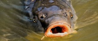
AEROMONOSIS In overgrown or enclosed ponds, less often in rivers and reservoirs, carp, carp, crucian carp, roach, rudd, bream, tench and even sturgeon affected by rubella (aeromonosis) are found. This is an infectious disease of fish.
Sick fish float on the surface of the reservoir without responding to external stimuli.
You can easily catch it with your hands. It looks like a watery, bloated monster:
its scales are ruffled, its eyes are bulging, and round red wounds and welts are often visible on its body.
You won’t put this kind of fish in a frying pan, and you won’t even want to pick it up.
And yet, if possible, it is necessary to catch it and bury it away from the reservoir, and disinfect it with lime milk.
LERNEOSIS In the hot season, crucian carp, tench, pike, burbot, carp, and silver carp can be affected by a parasitic crustacean. The disease caused by it is called Lerneosis. Abscess tubercles appear on the body of the fish, from the center of which an elastic greenish-brown rod emerges, a little more than a centimeter long. If you pull out this rod, at the hidden end you can see four glassy processes resembling the antlers of a deer. This is a parasitic crustacean that invades the body of the fish PISCICOLOSIS Sometimes a fisherman, having caught a bream or roach, pays attention to the black spots scattered throughout the body of the fish. This is a widespread disease and is called post-diplostomatosis, or black spot disease. (description above in the text) Caused by helminths carried by herons. In autumn, it is quite common to encounter carp, carp or tench, in which a mass of small, thin leeches with a spindle-shaped body covered with dark and white stripes is found in the head and front part of the body. One end of the leech - piscicola - has a small round suction cup, the other - a large spherical one. The disease caused by them is called piscicolosis. The disease is widespread in the lower reaches of the Volga and Don in the southern regions of the country.
SAPROLEGNIOSIS Fish restlessly swim around the pond, rub against piles, reeds, boats, are thrown out of the water, lose weight and react weakly to external stimuli. After the leech falls off, round wounds remain on the fish’s body, which often bleed. Various bacteria and fungi settle in places where the skin is damaged. Saprolegniosis is a fungal disease of fish caused by the mold Saprolegnia.
Fungi live in any water, are saprophytes and instantly settle on injured areas of the fish’s body and fish eggs.
They develop at any time of the year and even at low temperatures. A cotton-like coating appears on the fish’s body, fins, gills, and nasal openings. Penetrating into tissues, the fungus destroys them. Often, after catching a sick fish, you can find that it does not have a tail or pectoral fin. Over time, Saprolegnia penetrates the muscles and internal organs. Severely affected fish weaken and die. If you catch such a fish, do not rush to throw it into the pond; it is better to give it to birds or other animals as food. ARGULOSIS In thickets of reeds, reeds, cattails, pondweed, elodea and water buckwheat, in weakly flowing reservoirs, carp and perch are sometimes found, on the surface of which small (from 5 to 12 millimeters) mobile greenish crustaceans are visible.
This parasite has a spiny proboscis with which it pierces the skin of fish, feeding on their blood. Small wounds form at the site of damage. The disease caused by the parasite is called argulosis. Crustaceans swim freely in water, so they are easily transferred from pond to pond; the carrier may also be infected baitfish. CARP POX In the middle zone and in the south of the country, you often come across young carps with dense paraffin-shaped growths on the body. This is carp pox, a viral disease that is dangerous only for carp.
PIKE PLAGUE In autumn, during the pre-winter feast of predatory fish, the snout of a caught pike appears to be smeared with bright red lipstick. These are subcutaneous hemorrhages. They are also visible on the body near the pectoral fins. Often crescent-shaped wounds appear on the body. We're dealing with a plague here. In addition to pike, perch, burbot, bream and roach suffer from the plague. The disease was first reported in Germany before the Second World War. After the war, due to uncontrolled transportation of pike for fish farming purposes, diseased fish ended up in water bodies in the Moscow region and other areas. Now the plague of pikes is observed in the reservoirs of Northern Kazakhstan and the central zone of the country. It is not dangerous for humans.
However, poor heat treatment of sick fish can cause food poisoning.
Lerneosis, post-diplostomatoe, piscicolosis, saprolegniosis, argulosis affect only the surface of the fish’s body. Therefore, after thorough cleaning, such fish can be eaten without restrictions.
PHILOMETROIDOSES Many people, when cleaning carp under the scales of fish, find pinkish-red helminths rolled into a ring, up to 10-12 cm long, up to 1 mm thick.
These are phylometroidosis - round dioecious worms.
The females are located in the scale pockets of the carp, and the males are usually localized in the walls of the swim bladder. Males are much smaller than females, only 3-4 mm long. Females mature in the spring and release several hundred thousand small living larvae into the water. These larvae are eaten by cyclops, and through them fish become infected with phylometroidosis. The development cycle of these helminths is completed within a year. For humans, fish with phylometroidosis are not dangerous.
Helminths are removed when cleaning fish, and besides, they cannot exist in the human body.
But this disease causes significant harm to the fish itself. When infestation is high, juvenile carp die and the fish grow slowly. Saprolegnia develops in areas of the skin affected by helminths, and the fish loses its marketable appearance. Phylometroidosis of carp is being actively combated in pond farms.
Many fish farms have been completely freed from this disease. In commercial fish, such as crucian carp, bream, and silver bream, phylometroidosis can also sometimes be found. Adult helminths that parasitize crucian carp are usually found in the caudal or dorsal fins; the larvae infect the internal organs of the fish.
DIPLOSTOMIASIS Among silver carp, peled, trout and many other species of fish, sometimes you come across specimens that have cloudy eyes with an opaque lens. The reason for this is most often the larvae of small digenetic flukes that parasitize the lens and vitreous body of the eye. Sexually mature helminths live in the intestines of fish-eating birds. Birds become infected by eating diseased fish, which, due to their blindness, become easy prey for them. From helminth eggs that fall into the water with bird excrement, larvae emerge - miracidia, which penetrate the body of mollusks.
After a complex cycle of asexual development in the body of mollusks, numerous cercariae emerge into the water and attack fish.
Once in the fish, the cercariae penetrate the lens and transform into metacercariae, which can cause infection in birds. These helminths are not dangerous for humans.
However, the fish goes blind and grows worse.
Juvenile fish can die from the cercarial form of diplostomosis, when a mass of cercariae penetrates the skin into the fish’s body. DISEASE PREVENTION In the vast majority of cases, human infection occurs by eating raw, undercooked and undercooked fish. Thorough cooking is a reliable way to prevent diseases caused by fish.
For example,
tapeworm
larvae be
found in both lake and sea fish.
If you treat the fish appropriately, the tapeworm larvae will die. Tapeworm larvae die
at a temperature of +55 °C, i.e., if you boil or fry the fish in the usual way, the larvae will be killed.
The cooking time should be at least 10-15 minutes. A reliable way to avoid infection is to freeze the fish.
Fish weighing less than one kilogram is frozen in ordinary home freezers in 10 hours.
In large fish, tapeworm larvae are destroyed if they are kept in the freezer for one day. In other cases, it is recommended to disinfect fish by deep freezing.
At a temperature of -20 °C, fish must be kept for at least 3 days, at a temperature of -8...-10 °C - for 3-4 weeks.
The larvae also die with excessive salting.
Salt must be taken in an amount of at least 12% of the weight of the fish.
You need to keep the fish in salt for at least 5 days before eating it. When frying, the size of the pieces of fish is more important than their weight.
Small fish must be fried for at least 10 minutes.
Fish weighing 700-1200 g or fillets 2-3 cm thick are fried for 15-20 minutes. Pieces of fish thicker than 6 cm, as well as large uncut fish that have not had their backbone removed, should be fried for at least 40 minutes. Hot smoked fish, as well as carefully baked over an open fire, are harmless. smoke fish (cold and hot)
caught from a reservoir where there is a disease, since at home it is difficult to achieve uniform and sufficiently deep heating of fish, which is obtained at industrial fish processing plants.
You should not try to determine on your own whether the fish is infected or not.
Isolation of small parasite cysts requires special equipment and can only be carried out by specialists.
It is prohibited to feed raw fish from unfavorable water bodies to animals. After cutting the fish, the insides and scales must be destroyed, preventing them from being eaten by domestic or wild animals. This simple measure helps stop the spread of diseases to other bodies of water.
Treatment of helminthic diseases should be carried out only under the guidance of a doctor. Self-medication is unacceptable.
It should be remembered that alcohol does not play a preventive role in helminthiasis in the slightest degree. Surrounded by cysts and dense shells, helminths and their larvae have a very advanced enzymatic defense mechanism and withstand the action of digestive juices, resist even many potent substances, not to mention alcohol solutions. In this regard, it is prohibited to drink alcohol while fishing.
An intoxicated person loses control over his actions, loses his sense of vigilance, and the danger of infection increases many times over.
FOOD POISONING When eating foods containing toxic substances of bacterial or non-bacterial origin, food poisoning occurs. They can also become a source of poisoning. The caviar of marinkas, osmans, and some longhorned beetles that live in our reservoirs is poisonous. When marinka caviar is boiled, its toxic properties disappear. In case of poisoning by these fish,
vomiting, dizziness, weakness, and abdominal pain are noted.
In this case, it is recommended to do a gastric lavage, give a saline laxative, as well as a warm solution of potassium permanganate (1:1000, i.e. 1 g per 1 liter of water). It is imperative
that when the first signs of poisoning appear or if you suspect poisoning, you should immediately consult a doctor.
There is information
that during spawning the milt, caviar, and liver of burbot, pike, barbel, perch, beluga, mackerel, and haddock are poisonous.
The source of poisoning can be the mucus of lampreys. To remove it, lampreys are generously sprinkled with salt, after which the mucus is removed with a stiff brush. FOOD TOXIC INFECTIONS Food toxic infections occur when eating products, including fish, contaminated with certain types of pathogens: salmonella, colibacteria, proteus, staphylococci. The source of infection is domestic animals and poultry. A sick or healthy person who carries the bacteria can also be a source of infection. A reliable preventive measure against food poisoning is compliance with sanitary and hygienic requirements. Source: Newspaper “Fisherman of Fisherman” Authors: L. Grechanichenko; Sergey Grach. Photo from the network. Compiled by: V.A. Prokofiev